Pneumoperitoneum
Pneumoperitoneum is pneumatosis (abnormal presence of air or other gas) in the peritoneal cavity, a potential space within the abdominal cavity. The most common cause is a perforated abdominal organ, generally from a perforated peptic ulcer, although any part of the bowel may perforate from a benign ulcer, tumor or abdominal trauma. A perforated appendix seldom causes a pneumoperitoneum.
| Pneumoperitoneum | |
|---|---|
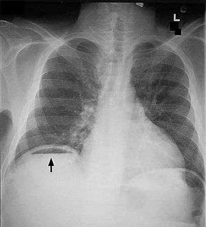 | |
| Frontal chest X-ray. The air bubble below the right hemidiaphragm (on the left of the image) is a pneumoperitoneum. | |
| Specialty | Gastroenterology |
A spontaneous pneumoperitoneum is a rare case that is not caused by an abdominal organ rupture. This is also called an idiopathic spontaneous pneumoperitoneum when the cause is not known.
In the mid-twentieth century, an "artificial" pneumoperitoneum was sometimes intentionally administered as a treatment for a hiatal hernia. This was achieved by insufflating the abdomen with carbon dioxide. The practice is currently used by surgical teams in order to perform laparoscopic surgery.
Causes
- Perforated duodenal ulcer – The most common cause of rupture in the abdomen. Especially of the anterior aspect of the first part of the duodenum.
- Perforated peptic ulcer
- Bowel obstruction
- Ruptured diverticulum
- Penetrating trauma
- Ruptured inflammatory bowel disease (e.g., megacolon)
- Necrotising enterocolitis/pneumatosis coli[1]
- Bowel cancer
- Ischemic bowel
- Steroids
- After laparotomy
- After laparoscopy
- Breakdown of a surgical anastomosis
- Bowel injury after endoscopy
- Peritoneal dialysis (PD), although the prevalence of pneumoperitoneum is estimated to be less than 4% among people with PD in a more recent study in the United Kingdom.[2]
- Vaginal insufflation (air enters via the fallopian tubes; e.g., water-skiing,[3] oral sex[4][5])
- Colonic or peritoneal infection
- From chest (e.g., bronchopleural fistula)
- Non-invasive PAP (positive airway pressure) can force air down duodenum as well as down trachea.
Spontaneous pneumoperitoneum
A spontaneous pneumoperitoneum is a rare case that is not caused by an abdominal organ rupture. This is also called an idiopathic spontaneous pneumoperitoneum when the cause is not known.[6] Causes of a spontaneous pneumoperitoneum, with no peritonitis include a barotrauma due to mechanical ventilation, and a tracheal rupture following an emergency intubation. In the ventilation case, air had passed from the chest into the abdominal cavity through the diaphragm. In the tracheal rupture air had passed along the great vessels.[7]
Diagnosis
When present, pneumoperitoneum can often be seen on projectional radiography, but small amounts are often missed, and CT scan is nowadays regarded as a criterion standard in the assessment of a pneumoperitoneum.[8] CT can visualize quantities as small as 5 cm³ of air or gas.
Signs that can be seen on projectional radiography are the double wall sign (also called Rigler's sign) and the football sign.
The double wall sign marks the presence of air on both sides of the intestine.[9] However, a false double wall sign can result from two loops of bowel being in contact with one another.[10] The sign is named after Leo George Rigler. It is not the same as Rigler's triad.
The football sign is when the abdomen appears as a large oval radiolucency reminiscent of an American football on a supine projectional radiograph.[11] The football sign is most frequently seen in infants with spontaneous or iatrogenic gastric perforation causing pneumoperitoneum. It is also seen in bowel obstruction with secondary perforation, as in Hirschprung disease, midgut volvulus, meconium ileus and intestinal atresia. Iatrogenic causes like endoscopic perforation may also give football sign.
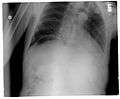 Another pneumoperitoneum on chest X-ray.
 Pneumoperitoneum seen on X-ray with the patient lying on his left side.
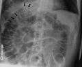 Double wall sign. This is a secondary sign of pneumoperitoneum. Patient is supine, and air within the abdomen and lumen of the bowel accentuate both sides of the bowel wall.
- Ultrasound finding of pneumoperitoneum known as "peritoneal stripe sign"[12]
Differential diagnosis
As differential diagnoses, a subphrenic abscess, bowel interposed between diaphragm and liver (Chilaiditi syndrome), and linear atelectasis at the base of the lungs can simulate free air under the diaphragm on a chest X-ray.
Treatment
Depends on causes
Usually a surgical consultation is indicated.
Terminology
Pneumoperitoneum can be described as peritoneal emphysema,[13] just as pneumomediastinum can be called mediastinal emphysema, but pneumoperitoneum is the usual name.
References
- Necrotizing Enterocolitis Bugs, Drugs and Things That Go Bump in the Night
- Imran, M.; Bhat, R.; Anijeet, H. (2011). "Pneumoperitoneum in peritoneal dialysis patients; one centre's experience". Clinical Kidney Journal. 4 (2): 120–123. doi:10.1093/ndtplus/sfq208. ISSN 2048-8505. PMC 4421564. PMID 25984130.
- Kizer, KW (May 1980). "Medical hazards of the water skiing douche". Ann Emerg Med. 9 (5): 268–269. doi:10.1016/s0196-0644(80)80387-8. PMID 7369581.
Many injuries can result from water skiing accidents, but the water skiing "douche" is unique to this sport. Although generally causing only discomfort, significant trauma can result from the forceful entry of water into the various body orifices.
- Jacobs VR, Mundhenke C, Maass N, Hilpert F, Jonat W (Oct 2000). "Sexual activity as cause for non-surgical pneumoperitoneum". JSLS. 4 (4): 297–300. PMC 3113190. PMID 11051188.
- Cawich SO, Johnson PB, Williams E, Naraynsingh V (25 Sep 2013). "Non-surgical pneumoperitoneum after oro-genital intercourse". Int J Surg Case Rep. 4 (11): 1048–1051. doi:10.1016/j.ijscr.2013.08.022. PMC 3825970. PMID 24121052.
- Pfister, J (September 1993). "Idiopathic spontaneous pneumoperitoneum. Case discussion based on two cases, assessment procedure and therapy and review of the literature". Helv Chir Acta. 60 (1–2): 49–56. PMID 8226082.
- Gutkin, Z (July 1992). "Spontaneous pneumoperitoneum without peritonitis". Int Surg. 77 (3): 219–23. PMID 1399374.
- Ali Nawaz Khan (2017-12-06). "eMedicine.com: Pneumoperitoneum". Cite journal requires
|journal=(help) - Harkin DW, Blake G. Small bowel obstruction in a young adult. Postgrad Med J. 1999 Mar;75(881):173-5. PMID 10448501. Free Full Text.
- de Lacey G, Bloomberg T, Wignall BK. Pneumoperitoneum: the misleading double wall sign. Clin Radiol. 1977 Jul;28(4):445-8. PMID 872511.
- Rampton JW (April 2004). "The football sign". Radiology. 231 (1): 81–2. doi:10.1148/radiol.2311011290. PMID 14990817.
- "UOTW #68 - Ultrasound of the Week". Ultrasound of the Week. 22 March 2016. Retrieved 27 May 2017.
- Marian, K; et al. (2015), "Spontaneous pneumoperitoneum in a patient after ventilation therapy", Pol Przegl Chir, 86 (12): 601–603, doi:10.1515/pjs-2015-0008, PMID 25803061.
External links
| Classification | |
|---|---|
| External resources |