Adenomyomatosis
Adenomyomatosis is a benign condition characterized by hyperplastic changes of unknown cause involving the wall of the gallbladder.[1] Adenomyomatosis is caused by an overgrowth of the mucosa, thickening of the muscular wall, and formation of intramural diverticula or sinus tracts termed Rokitansky–Aschoff sinuses, also called entrapped epithelial crypts.
| Adenomyomatosis | |
|---|---|
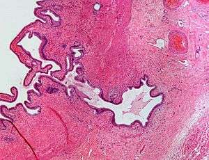 | |
| Micrograph showing Rokitansky–Aschoff sinus. H&E stain. |
Pathophysiology
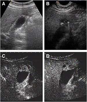
a The fundus of the gallbladder wall was thickened and the GB wall was obscure.
b The intramural echogenic foci were detected by high frequency transducer.
c CEUS—arterial phase (22 s) —heterogeneous hyper-enhancement and wall was intact.
d CEUS—venous phase (34 s) the anechoic spaces were more clear.
Rokitansky–Aschoff sinuses
Rokitansky–Aschoff sinuses are pseudodiverticula or pockets in the wall of the gallbladder. They may be microscopic or macroscopic. Histologically, they are outpouchings of gallbladder mucosa into the gallbladder muscle layer and subserosal tissue as a result of hyperplasia and herniation of epithelial cells through the fibromuscular layer of the gallbladder wall.[4]
Rokitansky–Aschoff sinuses are not of themselves considered abnormal but they can be associated with cholecystitis.[5]
They form as a result of increased pressure in the gallbladder and recurrent damage to the wall of the gallbladder.[6]
Associations
Black pigment gallstones can form in Rokitansky–Aschoff sinuses of the gallbladder after the fourth to fifth decades of life in absence of the typical risk factors for bilirubin suprasaturation of bile.[4] Hence, they are associated with gallstones (cholelithiasis). Cases of gall bladder cancer have also been reported to arise from Rokitansky–Aschoff sinuses.[7]
Diagnosis
On abdominal ultrasound, Rokitansky–Aschoff sinuses are seen as highly echogenic nodes with "comet tail" reverberation, which represent cholesterol crystals.[8] This finding is very specific for adenomyomatosis.[8] Magnetic resonance imaging also plays an important role in the diagnosis of Rokitansky–Aschoff sinuses.[9]
Eponym
Rokitansky–Aschoff sinuses are named after Carl Freiherr von Rokitansky (1804–1878), a pathologist in Vienna, Austria and Ludwig Aschoff (1866–1942), a pathologist in Bonn, Germany.[10][11]
References
- Ram and Midha (August 1975). "Adenomyomatosis of the gallbladder". Surgery. 78 (2): 224–229. PMID 1154265.
- "UOTW #79 - Ultrasound of the Week". Ultrasound of the Week. 16 April 2017. Retrieved 27 May 2017.
- Tang, ShaoShan; Huang, LiPing; Wang, Yao; Wang, YiJiao (2015). "Contrast-enhanced ultrasonography diagnosis of fundal localized type of gallbladder adenomyomatosis". BMC Gastroenterology. 15 (1): 99. doi:10.1186/s12876-015-0326-y. ISSN 1471-230X. PMC 4524444. PMID 26239485.
- Cariati, A; Cetta, F (2002). "Rokitansky-Aschoff sinuses of the gallbladder are associated with black pigment gallstone formation: a scanning electron microscopy study". Ultrastructural Pathology. 27 (4): 265–70. doi:10.1080/01913120309913. PMID 12907372.
- van Breda Vriesman, AC; Engelbrecht, MR; Smithuis, RH; Puylaert, JB (February 2007). "Diffuse gallbladder wall thickening: differential diagnosis". AJR. American Journal of Roentgenology. 188 (2): 495–501. doi:10.2214/AJR.05.1712. PMID 17242260.
- Stunell, H; Buckley, O; Geoghegan, T; O’Brien, J; Ward, E; Torreggiani, W (2008). "Imaging of adenomyomatosis of the gall bladder". Journal of Medical Imaging and Radiation Oncology. 52 (2): 109–117. doi:10.1111/j.1440-1673.2008.01926.x. ISSN 1754-9477. PMID 18373800.
- Matsumoto, T; Shimada, K (November 2009). "A case of gallbladder cancer arising from the Rokitansky-Aschoff sinus". Japanese Journal of Clinical Oncology. 39 (11): 776. doi:10.1093/jjco/hyp149. PMID 19884193.
- Ayush Goel; Yoshiharu Ryu; et al. "Adenomyomatosis of the gallbladder". Radiopaedia. Retrieved 2018-02-01.
- Yoshimitsu, K; Honda, H; Jimi, M; Kuroiwa, T; Hanada, K; Irie, H; Tajima, T; Takashima, M; Chijiiwa, K; Shimada, M; Masuda, K (June 1999). "MR diagnosis of adenomyomatosis of the gallbladder and differentiation from gallbladder carcinoma: importance of showing Rokitansky-Aschoff sinuses". AJR. American Journal of Roentgenology. 172 (6): 1535–40. doi:10.2214/ajr.172.6.10350285. PMID 10350285.
- synd/983 at Who Named It?
- Kanne, JP; Rohrmann CA, Jr; Lichtenstein, JE (2005). "Eponyms in radiology of the digestive tract: historical perspectives and imaging appearances. Part 2. Liver, biliary system, pancreas, peritoneum, and systemic disease". Radiographics. 26 (2): 465–80. doi:10.1148/rg.262055130. PMID 16549610.