Intestinal atresia
Intestinal atresia is any congenital malformation of the intestine which causes bowel obstruction. The malformation can be a narrowing (stenosis), absence, or rotation of a portion of the intestine. These defects can either occur in the small or large intestine.
| Intestinal atresia | |
|---|---|
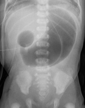 | |
| Radiograph with double bubble sign indicating duodenal atresia | |
| Specialty | Gastroenterology |
Types
By location
Different types of intestinal atresia may be classified by their location. Patients may have intestinal atresia in multiple locations.[1]
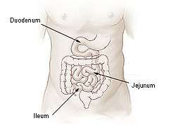
- Duodenal atresia – malformation of the duodenum, part of the intestine that empties from the stomach, and first section of the small intestine
- Jejunal atresia – malformation of the jejunum, the second part of the small intestine extending from the duodenum to the ileum, that causes the jejunum to block blood flow to the colon [2]
- Ileal atresia – malformation of the ileum, the lower part of the small intestine
- Colon atresia – malformation of the colon
Malformations may also occur along multiple portions of the intestinal tract; for instance a malformation that occurs along or spans the length of the jejunum and the ilieum is termed jejunoileal atresia.[3][4]
By malformation
Intestinal atresia can also by classified by the type of malformation.[5] The classification system by Louw and Barnard (1955) divided them into four families of malformation:[6][5][3]
- Type I – web with complete or incomplete obstruction
- Type II – blind end (complete obstruction)
- Type III – flaws in the blood supply
- Type IIIa – mesenteric gap
- Type IIIb – apple peel syndrome
- Type IV – multiple blockages
In rare cases, the small intestine wraps around its vascular supply and forms a spiral; This is called apple peel bowel or apple-peel intestinal atresia if it occurs along the jejunum, Christmas tree intestinal atresia if it occurs along the duodenum,[7][8] and may occur along both.[1]
Cause
The most common cause of non-duodenal intestinal atresia is a vascular accident in utero that leads to decreased intestinal perfusion and ischemia of the respective segment of bowel.[4] This leads to narrowing, or in the most severe cases, complete obliteration of the intestinal lumen.
In the case that the superior mesenteric artery, or another major intestinal artery, is occluded, large segments of bowel can be entirely underdeveloped (Type III). Classically, the affected area of bowel assumes a spiral configuration and is described to have an "apple peel" like appearance; this is accompanied by lack of a dorsal mesentery (Type IIIb).
An inherited form – familial multiple intestinal atresia – has also been described. This disorder was first reported in 1971.[9] It is due to a mutation in the gene TTC7A on short arm of chromosome 2 (2p16). It is inherited as an autosomal recessive gene and is usually fatal in infancy.
Ileal atresia can also result as a complication of meconium ileus.
Epidemiology
The most common form of intestinal atresia is duodenal atresia. It has a strong association with Down syndrome.[10] The second most common type is ileal atresia.
Prevalence of jejunoileal atresia is 1 to 3 in 10,000 live births. It is weakly associated with cystic fibrosis, intestinal malrotation, and gastroschisis.[4]
Diagnosis
Intestinal atresias are often discovered before birth; either during a routine sonogram which shows a dilated intestinal segment due to the blockage, or by the development of polyhydramnios (the buildup of too much amniotic fluid in the uterus). These abnormalities are indications that the fetus may have a bowel obstruction which a more detailed ultrasound study can confirm.[3]
Some fetuses with bowel obstruction have abnormal chromosomes. An amniocentesis is recommended because it can determine not only the sex of the baby, but whether or not there is a problem with the chromosomes.
If not diagnosed in utero, infants with intestinal atresia are typically diagnosed at day 1 or day 2 after presenting with eating problems, vomiting, and/or failure to have a bowel movement.[4] Diagnosis can be confirmed with an X-ray, and typically followed with an upper gastrointestinal series, lower gastrointestinal series, and ultrasound.[3][4]
Complications can include rupture of the bowel before or after birth.
Treatment
Fetal and neonatal intestinal atresia are treated using laparotomy after birth. If the area affected is small, the surgeon may be able to remove the damaged portion and join the intestine back together. In instances where the narrowing is longer, or the area is damaged and cannot be used for period of time, a temporary stoma may be placed.
See also
References
- Federici, S.; Domenichelli, V.; Antonellini, C.; Dòmini, R. (August 2003). "Multiple intestinal atresia with apple peel syndrome: successful treatment by five end-to-end anastomoses, jejunostomy, and transanastomotic silicone stent". Journal of Pediatric Surgery. 38 (8): 1250–1252. doi:10.1016/S0022-3468(03)00281-1. ISSN 1531-5037. PMID 12891506.
- Jejunal Atresia. Genetic and Rare Diseases Information Center (GARD) https://rarediseases.info.nih.gov/diseases/6799/jejunal-atresia
- "Jejunoileal Atresia". Short Bowel Foundation. Retrieved 2019-02-03.
- William J. Cochran, MD. "Jejunoileal Atresia - Pediatrics". MSD Manual Professional Edition. Retrieved 2019-02-03.
- Shorter, Nicholas A.; Georges, Anthony; Perenyi, Agnes; Garrow, Eugene (Nov 2006). "A proposed classification system for familial intestinal atresia and its relevance to the understanding of the etiology of jejunoileal atresia". Journal of Pediatric Surgery. 41 (11): 1822–1825. doi:10.1016/j.jpedsurg.2006.06.008. ISSN 0022-3468. PMID 17101351.
- Louw, J.H.; Barnard, C.N. (Nov 1955). "CONGENITAL INTESTINAL ATRESIA OBSERVATIONS ON ITS ORIGIN". The Lancet. 266 (6899): 1065–1067. doi:10.1016/s0140-6736(55)92852-x. ISSN 0140-6736.
- Blyth, H; Dickson, J A (September 1969). "Apple peel syndrome (congenital intestinal atresia): a family study of seven index patients". Journal of Medical Genetics. 6 (3): 275–277. doi:10.1136/jmg.6.3.275. ISSN 0022-2593. PMC 1468754. PMID 5345098.
- Gaillard, Frank. "Apple-peel intestinal atresia". Radiopaedia. Retrieved 2019-02-01.
- Mishalany, Henry G.; Der Kaloustian, Vazken M. (July 1971). "Familial multiple-level intestinal atresias: Report of two siblings". The Journal of Pediatrics. 79 (1): 124–125. doi:10.1016/s0022-3476(71)80072-0. ISSN 0022-3476.
- Le, Tao (2013). First Aid for the USMLE STEP. New York: Mc Graw Hill. p. 308. ISBN 978-0-07-180232-1.
External links
| Classification |
|---|