Parasympathetic root of ciliary ganglion
The parasympathetic root of ciliary ganglion provides parasympathetic innervation to the ciliary ganglion.
| Parasympathetic root of ciliary ganglion | |
|---|---|
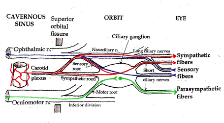 Pathways in the Ciliary Ganglion. Green = parasympathetic; Red = sympathetic; Blue = sensory | |
| Details | |
| From | Edinger-Westphal nucleus |
| To | short ciliary nerves |
| Identifiers | |
| Latin | ramus parasympathica ganglii ciliaris |
| TA | A14.2.01.010 |
| FMA | 52578 |
| Anatomical terms of neuroanatomy | |
The ciliary ganglion is a parasympathetic ganglion. Incoming parasympathetic nerve fibers form synapses with the dendrites of nerve cells within the ganglion. However, the ciliary ganglion is not simply a relay station connecting preganglionic to postganglionic nerve fibers. There are roughly twice as many incoming parasympathetic fibers as outgoing parasympathetic fibers. Neural processing occurs as incoming signals converge onto target neurons.
Presynaptic
Presynaptic parasympathetic fibers originate in the Edinger-Westphal nucleus, the parasympathetic motor nucleus associated with the oculomotor nucleus in the brainstem. Axons from the Edinger-Westphal nucleus and the oculomotor nucleus run together in the brainstem and exit together as the oculomotor nerve (cranial nerve III). The oculomotor nerve passes through the lateral wall of the cavernous sinus and enters the orbit through the superior orbital fissure. It divides into branches that innervate the levator palpebrae superioris and four of the six extraocular muscles. Parasympathetic fibers initially run in the inferior division of the oculomotor nerve. They exit as one or two short “motor roots” that synapse in the ciliary ganglion.
Postsynaptic
Postsynaptic parasympathetic fibers leave the ciliary ganglion in multiple (six to ten) short ciliary nerves. These nerves enter the posterior aspect of the eyeball to innervate the sphincter pupillae and ciliaris muscles. The sphincter pupillae constricts the iris. The ciliaris muscle changes the shape of the lens, allowing the eye to focus on nearby objects (accommodation). Neither of these muscles is under voluntary control.
External links
Anatomy of the autonomic nervous system | |||||
|---|---|---|---|---|---|
| Head |
| ||||
| Neck |
| ||||
| Chest |
| ||||
| Abdomen |
| ||||
| Pelvis |
| ||||
| |||||
Optical illusions (list) | ||
|---|---|---|
| Illusions |
| |
| Popular culture |
| |
| Related |
| |
| Authority control |
|---|