Long ciliary nerves
The long ciliary nerves, two or three in number, are given off from the nasociliary nerve as it crosses the optic nerve. The nasociliary nerve that the long ciliary nerves branch from is itself a branch of the ophthalmic branch (V1) of the trigeminal nerve (CN V).
| Long ciliary nerves | |
|---|---|
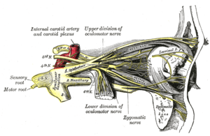 Nerves of the orbit, and the ciliary ganglion. Side view. | |
| Details | |
| From | Nasociliary nerve |
| Fiber type | "Somatosensory" (via V1 Lacrimal), and "Sympathetic" (via V2 Zygomatic) |
| Identifiers | |
| Latin | nervi ciliares longi |
| TA | A14.2.01.027 |
| FMA | 52691 |
| Anatomical terms of neuroanatomy | |
They accompany the short ciliary nerves from the ciliary ganglion, pierce the posterior part of the sclera, and running forward between it and the choroid, are distributed to the iris and cornea.
The long ciliary nerves provide sensory innervation to the eyeball, including the cornea. In addition, they contain sympathetic fibers from the superior cervical ganglion to the dilator pupillae muscle. The sympathetic fibers to the dilator pupillae muscle mainly travel in the nasociliary nerve but there are also sympathetic fibers in the short ciliary nerves that pass through the ciliary ganglion without forming synapses.
See also
Additional images
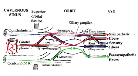 Pathways in the Ciliary Ganglion.
References
This article incorporates text in the public domain from page 888 of the 20th edition of Gray's Anatomy (1918)