Stylomastoid foramen
Between the styloid and mastoid processes of the temporal bone is the stylomastoid foramen
| Stylomastoid foramen | |
|---|---|
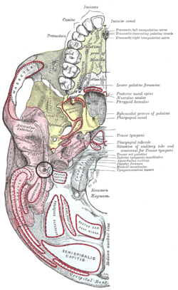 Base of skull. Inferior surface. Pink region is temporal bone, and stylomastoid foramen is in black circle at center of pink region. | |
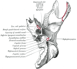 Left temporal bone. Inferior surface. (Stylomastoid foramen is third label from the bottom on the left.) | |
| Details | |
| Identifiers | |
| Latin | foramen stylomastoideum |
| TA | A02.1.06.048 |
| FMA | 55816 |
| Anatomical terms of bone | |
It is the termination of the facial canal, and transmits the facial nerve and stylomastoid artery.
Clinical relevance
Bell's palsy can result from inflammation of the facial nerve where it leaves the skull at the stylomastoid foramen. Patients with Bell's palsy appear with facial drooping on the affected side.
Additional images
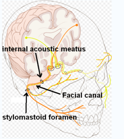 Facial canal
 Lateral head anatomy detail.Facial nerve dissection.
References
This article incorporates text in the public domain from page 144 of the 20th edition of Gray's Anatomy (1918)
External links
- Anatomy figure: 22:4a-07 at Human Anatomy Online, SUNY Downstate Medical Center
- Anatomy photo:22:os-0708 at the SUNY Downstate Medical Center
- "Anatomy diagram: 34257.000-1". Roche Lexicon – illustrated navigator. Elsevier. Archived from the original on 2014-01-01.
- lesson3 at The Anatomy Lesson by Wesley Norman (Georgetown University) (midearcavity)
- Diagram at patientsforum.com
| Authority control |
|---|
This article is issued from
Wikipedia.
The text is licensed under Creative
Commons - Attribution - Sharealike.
Additional terms may apply for the media files.