Abdomen
The abdomen (colloquially called the belly,[1] tummy or midriff) is the part of the body between the thorax (chest) and pelvis, in humans and in other vertebrates. The abdomen is the front part of the abdominal segment of the trunk. The region occupied by the abdomen is called the abdominal cavity. In arthropods it is the posterior tagma of the body; it follows the thorax or cephalothorax.[2][3]
| Abdomen | |
|---|---|
 | |
 The human abdomen and organs which can be found beneath the surface | |
| Details | |
| Actions | Movement and support for the torso Assistance with breathing Protection for the inner organs Postural support |
| Identifiers | |
| Latin | Abdomen |
| MeSH | D000005 |
| TA | A01.1.00.016 |
| FMA | 9577 |
| Anatomical terminology | |
In humans, the abdomen stretches from the thorax at the thoracic diaphragm to the pelvis at the pelvic brim. The pelvic brim stretches from the lumbosacral joint (the intervertebral disc between L5 and S1) to the pubic symphysis and is the edge of the pelvic inlet. The space above this inlet and under the thoracic diaphragm is termed the abdominal cavity. The boundary of the abdominal cavity is the abdominal wall in the front and the peritoneal surface at the rear.
Structure
In vertebrates, the abdomen is a large body cavity enclosed by the abdominal muscles, at front and to the sides, and by part of the vertebral column at the back. Lower ribs can also enclose ventral and lateral walls. The abdominal cavity is continuous with, and above, the pelvic cavity. It is attached to the thoracic cavity by the diaphragm. Structures such as the aorta, inferior vena cava and esophagus pass through the diaphragm. Both the abdominal and pelvic cavities are lined by a serous membrane known as the parietal peritoneum. This membrane is continuous with the visceral peritoneum lining the organs.[4] The abdomen in vertebrates contains a number of organs belonging to, for instance, the digestive system, urinary system, and muscular system.
Contents
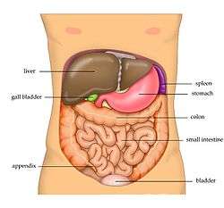
The abdominal cavity contains most organs of the digestive system, including the stomach, the small intestine, and the colon with its attached appendix. Other digestive organs are known as the accessory digestive organs and include the liver, its attached gallbladder, and the pancreas, and these communicate with the rest of the system via various ducts. The spleen, and organs of the urinary system including the kidneys, and adrenal glands also lie within the abdomen, along with many blood vessels including the aorta and inferior vena cava. The urinary bladder, uterus, fallopian tubes, and ovaries may be seen as either abdominal organs or as pelvic organs. Finally, the abdomen contains an extensive membrane called the peritoneum. A fold of peritoneum may completely cover certain organs, whereas it may cover only one side of organs that usually lie closer to the abdominal wall. This is called the retroperitoneum, and the kidneys and ureters are known as retroperitoneal organs.
Abdominal organs can be highly specialized in some animals. For example, the stomach of ruminants, (a suborder of mammals that includes cattle and sheep), is divided into four chambers – rumen, reticulum, omasum and abomasum.[5]
 View of the various organs and blood-vessels in proximity with liver.
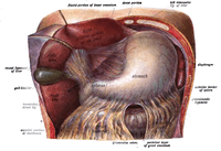 Liver lifted to show gall bladder and stomach in situ.
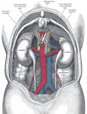 The relations of the viscera and large vessels of the abdomen, seen from behind.
Muscles

(Right) A male abdomen.
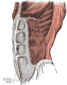
There are three layers of muscles in the abdominal wall. They are, from the outside to the inside: external oblique, internal oblique, and transverse abdominal.[6] The first three layers extend between the vertebral column, the lower ribs, the iliac crest and pubis of the hip. All of their fibers merge towards the midline and surround the rectus abdominis in a sheath before joining up on the opposite side at the linea alba. Strength is gained by the criss-crossing of fibers, such that the external oblique runs downward and forward, the internal oblique upward and forward, and the transverse abdominal horizontally forward.[6]
The transverse abdominal muscle is flat and triangular, with its fibers running horizontally. It lies between the internal oblique and the underlying transverse fascia. It originates from Poupart's ligament, the inner lip of the ilium, the lumbar fascia and the inner surface of the cartilages of the six lower ribs. It inserts into the linea alba behind the rectus abdominis.
The rectus abdominis muscles are long and flat. The muscle is crossed by three fibrous bands called the tendinous intersections. The rectus abdominis is enclosed in a thick sheath, formed as described above, by fibers from each of the three muscles of the lateral abdominal wall. They originate at the pubis bone, run up the abdomen on either side of the linea alba, and insert into the cartilages of the fifth, sixth, and seventh ribs. In the region of the groin, the inguinal canal, is a passage through the layers. This gap is where the testes can drop through the wall and where the fibrous cord from the uterus in the female runs. This is also where weakness can form, and cause inguinal hernias.[6]
The pyramidalis muscle is small and triangular. It is located in the lower abdomen in front of the rectus abdominis. It originates at the pubic bone and is inserted into the linea alba halfway up to the navel.
Function
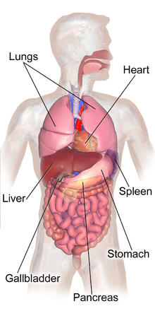
Functionally, the human abdomen is where most of the digestive tract is placed and so most of the absorption and digestion of food occurs here. The alimentary tract in the abdomen consists of the lower esophagus, the stomach, the duodenum, the jejunum, ileum, the cecum and the appendix, the ascending, transverse and descending colons, the sigmoid colon and the rectum. Other vital organs inside the abdomen include the liver, the kidneys, the pancreas and the spleen.
The abdominal wall is split into the posterior (back), lateral (sides), and anterior (front) walls.
Movement, breathing and other functions
The abdominal muscles have different important functions. They assist as muscles of exhalation in the breathing process during forceful exhalation. Moreover, these muscles serve as protection for the inner organs. Furthermore, together with the back muscles they provide postural support and are important in defining the form. When the glottis is closed and the thorax and pelvis are fixed, they are integral in the cough, urination, defecation, childbirth, vomit, and singing functions.[6] When the pelvis is fixed, they can initiate the movement of the trunk in a forward motion. They also prevent hyperextension. When the thorax is fixed, they can pull up the pelvis and finally, they can bend the vertebral column sideways and assist in the trunk's rotation.[6]
Posture
The transverse abdominis muscle is the deepest muscle, therefore, it cannot be touched from the outside. It can greatly affect the body's posture. The internal obliques are also deep and also affect body posture. Both of them are involved in rotation and lateral flexion of the spine and are used to bend and support the spine from the front. The external obliques are more superficial and they are also involved in rotation and lateral flexion of the spine. Also they stabilize the spine when upright. The rectus abdominis muscle is not the most superficial abdominal muscle. The tendonous sheath extending from the external obliques cover the rectus abdominis. The rectus abdominis is the muscle that very fit people develop into the 6-pack ab look. Although, it should really be a 10 pack as there are 5 vertical sections on each side. The 2 bottom sections are just above the pubic bone and usually not visible, hence, the 6 pack abs. The rectus abdominals' function is to bend one's back forward (flexion). The main work of the abdominal muscles is to bend the spine forward when contracting concentrically.[7]
Society and culture
Social and cultural perceptions of the outward appearance of the abdomen has varying significance around the world. Depending on the type of society, excess weight can be perceived as an indicator of wealth and prestige due to excess food, or as a sign of poor health due to lack of exercise. In many cultures, bare abdomens are distinctly sexualized and perceived similarly to breast cleavage.
Exercise
Being key elements of spinal support, and contributors to good posture, it is important to properly exercise the abdominal muscles together with the back muscles because when these are weak or overly tight they can suffer painful spasms and injuries. When properly exercised, abdominal muscles contribute to improved posture and balance, reduce the likelihood of back pain episodes, reduce the severity of back pain, protect against injury by responding efficiently to stresses, help avoid some back surgeries, and help with the healing of back problems, or after spine surgery. When strengthened, the abdominal muscles provide flexibility as well. The abdominal muscles can be worked by practicing disciplines of general body strength such as Pilates, yoga, T'ai chi, and jogging. There are also specific routines which target each of these muscles.
Clinical significance
Abdominal obesity is a condition where abdominal fat or visceral fat, has built up excessively between the abdominal organs. This is associated with a higher risk of heart disease, asthma and type 2 diabetes.
Abdominal trauma is an injury to the abdomen and can involve damage to the abdominal organs. There is an associated risk of severe blood loss and infection.[8] Injury to the lower chest can cause injuries to the spleen and liver.[9]
A scaphoid abdomen is when the abdomen is sucked inwards.[10] In a newborn, it may represent a diaphragmatic hernia.[11] In general, it is indicative of malnutrition.[12]
Disease
Many gastrointestinal diseases affect the abdominal organs. These include stomach disease, liver disease, pancreatic disease, gallbladder and bile duct disease; intestinal diseases include enteritis, coeliac disease, diverticulitis, and IBS.
Examination
Different medical procedures can be used to examine the organs of the gastrointestinal tract. These include endoscopy, colonoscopy, sigmoidoscopy, enteroscopy, oesophagogastroduodenoscopy and virtual colonoscopy. There are also a number of medical imaging techniques that can be used. Surface landmarks are important in the examination of the abdomen.
Surface landmarks
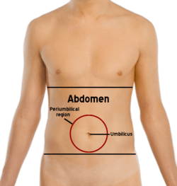
In the mid-line a slight furrow extends from the xiphoid process above to the pubic symphysis below, representing the linea alba in the abdominal wall. At about its midpoint sits the umbilicus or navel. The rectus abdominis on each side of the linea alba stands out in muscular people. The outline of these muscles is interrupted by three or more transverse depressions indicating the tendinous intersections. There is usually one about the xiphoid process, one at the navel, and one in between. It is the combination of the linea alba and the tendinous intersections which form the abdominal "six-pack" sought after by many people.
The upper lateral limit of the abdomen is the subcostal margin (at or near the subcostal plane) formed by the cartilages of the false ribs (8, 9, 10) joining one another. The lower lateral limit is the anterior crest of the ilium and Poupart's ligament, which runs from the anterior superior spine of the ilium to the spine of the pubis. These lower limits are marked by visible grooves. Just above the pubic spines on either side are the external abdominal rings, which are openings in the muscular wall of the abdomen through which the spermatic cord emerges in the male, and through which an inguinal hernia may rupture.
One method by which the location of the abdominal contents can be appreciated is to draw three horizontal and two vertical lines.
Horizontal lines
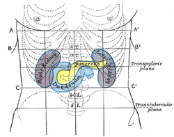
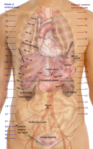
- The highest of the former is the transpyloric line of C. Addison, which is situated halfway between the suprasternal notch and the top of the pubic symphysis, and often cuts the pyloric opening of the stomach an inch to the right of the mid-line. The hilum of each kidney is a little below it, while its left end approximately touches the lower limit of the spleen. It corresponds to the first lumbar vertebra behind.
- The second line is the subcostal line, drawn from the lowest point of the subcostal arch (tenth rib). It corresponds to the upper part of the third lumbar vertebra, and it is an inch or so above the umbilicus. It indicates roughly the transverse colon, the lower ends of the kidneys, and the upper limit of the transverse (3rd) part of the duodenum.
- The third line is called the intertubercular line, and runs across between the two rough tubercles, which can be felt on the outer lip of the crest of the ilium about two and a half inches (64 mm) from the anterior superior spine. This line corresponds to the body of the fifth lumbar vertebra, and passes through or just above the ileo-caecal valve, where the small intestine joins the large intestine.
Vertical lines
The two vertical or mid-Poupart lines are drawn from the point midway between the anterior superior spine and the pubic symphysis on each side, vertically upward to the costal margin.
- The right one is the most valuable, as the ileo-caecal valve is situated where it cuts the intertubercular line. The orifice of the appendix lies an inch lower, at McBurney's point. In its upper part, the vertical line meets the transpyloric line at the lower margin of the ribs, usually the ninth, and here the gallbladder is situated.
- The left mid-Poupart line corresponds in its upper three-quarters to the inner edge of the descending colon.
The right subcostal margin corresponds to the lower limit of the liver, while the right nipple is about half an inch above its upper limit.
Quadrants and regions
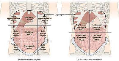
The abdomen can be divided into quadrants or regions to describe the location of an organ or structure. Classically, quadrants are described as the left upper, left lower, right upper, and right lower. Quadrants are also often used in describing the site of an abdominal pain.[13]
The abdomen can also be divided into nine regions.
| right hypochondriac/hypochondrium | epigastric/epigastrium | left hypochondriac/hypochondrium |
| right lumbar/flank/latus/lateral | umbilical | left lumbar/flank/latus/lateral |
| right inguinal/iliac | hypogastric/suprapubic | left inguinal/iliac |
These terms stem from "hypo" meaning "below" and "epi" means "above", while "chondron" means "cartilage" (in this case, the cartilage of the rib) and "gaster" means stomach. The reversal of "left" and "right" is intentional, because the anatomical designations reflect the patient's own right and left.)
The "right iliac fossa" (RIF) is a common site of pain and tenderness in patients who have appendicitis. The fossa is named for the underlying iliac fossa of the hip bone, and thus is somewhat imprecise. Most of the anatomical structures that will produce pain and tenderness in this region are not in fact in the concavity of the ileum. However, the term is in common usage.
Other animals
.jpg)
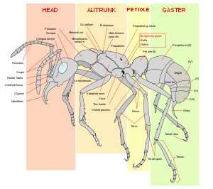
In arthropods the abdomen is built up of a series of upper plates known as tergites and lower plates known as sternites, the whole being held together by a tough yet stretchable membrane.
The abdomen contains the insect's digestive tract and reproductive organs, it consists of eleven segments in most orders of insects though the eleventh segment is absent in the adult of most higher orders. The number of these segments does vary from species to species with the number of segments visible reduced to only seven in the common honeybee. In the Collembola (Springtails) the abdomen has only six segments.
The abdomen is sometimes highly modified. In Apocrita (bees, ants and wasps), the first segment of the abdomen is fused to the thorax and is called the propodeum. In ants the second segment forms the narrow petiole. Some ants have an additional postpetiole segment, and the remaining segments form the bulbous gaster.[14] The petiole and gaster (abdominal segments 2 and onward) are collectively called the metasoma.
Unlike other arthropods, insects possess no legs on the abdomen in adult form, though the Protura do have rudimentary leg-like appendages on the first three abdominal segments, and Archaeognatha possess small, articulated "styli" which are sometimes considered to be rudimentary appendages. Many larval insects including the Lepidoptera and the Symphyta (Sawflies) have fleshy appendages called prolegs on their abdominal segments (as well as their more familiar thoracic legs), which allow them to grip onto the edges of plant leaves as they walk around.
In arachnids (spiders, scorpions and relatives), the term "abdomen" is used interchangeably with "opisthosoma" ("hind body"), which is the body section posterior to that bearing the legs and head (the prosoma or cephalothorax).
See also
References
- CHRISTINA STIEHL(12/21/2017). What Actually Causes a Beer Belly, and How to Get Rid of It. Thrillist. Access date [4 Feb 2019].
- Abdomen. (n.d.). Dictionary.com Unabridged (v 1.1). Retrieved 22 October 2007
- Abdomen. Dictionary.com. The American Heritage Dictionary of the English Language, 4th Edition. Retrieved 22 October 2007
- Peritoneum. The Veterinary Dictionary. Elsevier, 2007. Retrieved 22 October 2007
- "Ruminant." The Veterinary Dictionary. Elsevier, 2007. Retrieved 22 October 2007
- "Abdominal cavity". Encyclopædia Britannica. I: A-Ak – Bayes (15th ed.). Chicago, IL: Encyclopædia Britannica, Inc. 2010. pp. 19–20. ISBN 978-1-59339-837-8.
- "The Abdominal Muscle Group". Retrieved 2010-07-13.
- Jansen JO, Yule SR, Loudon MA (April 2008). "Investigation of blunt abdominal trauma". BMJ. 336 (7650): 938–42. doi:10.1136/bmj.39534.686192.80. PMC 2335258. PMID 18436949.
- Wyatt, Jonathon; Illingworth, RN; Graham, CA; Clancy, MJ; Robertson, CE (2006). Oxford Handbook of Emergency Medicine. Oxford University Press. p. 346. ISBN 978-0-19-920607-0.
- Dorland's illustrated medical dictionary (32nd ed.). Philadelphia: Saunders/Elsevier. 2012. p. 2. ISBN 978-1-4160-6257-8.
- Durward, Heather; Baston, Helen (2001). Examination of the newborn: a practical guide. New York: Routledge. p. 134. ISBN 978-0-415-19184-5.
- Ferguson, Charles (1990). "Chapter 93: Inspection, Auscultation, Palpation, and Percussion of the Abdomen". In Walker, HK; Hall, WD; JW, Hurst (eds.). Clinical Methods: The History, Physical, and Laboratory Examinations (3rd ed.). Boston: Butterworths. Retrieved 2013-11-27.
- Saladin, Kenneth (2011). Human anatomy. McGraw-Hill. p. 14. ISBN 9780071222075.
- "Glossary of Descriptive Terminology". Desertants.org. Archived from the original on 17 May 2013. Retrieved 2013-07-08.
External links
| Look up abdomen in Wiktionary, the free dictionary. |
| Wikimedia Commons has media related to Abdomen. |

| Authority control |
|
|---|