Iliac crest
The crest of the ilium (or iliac crest) is the superior border of the wing of ilium and the superiolateral margin of the greater pelvis.
| Iliac crest | |
|---|---|
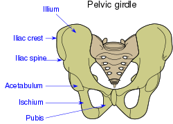 Pelvic girdle. | |
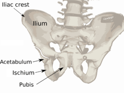 Overview of Ilium as largest bone of the pelvis. | |
| Details | |
| Identifiers | |
| Latin | crista iliaca |
| TA | A02.5.01.106 |
| FMA | 16914 |
| Anatomical terms of bone | |
Structure
The iliac crest stretches posteriorly from the anterior superior iliac spine (ASIS) to the posterior superior iliac spine (PSIS). Behind the ASIS, it divides into an outer and inner lip separated by the intermediate zone. The outer lip bulges laterally into the iliac tubercle. [1] Palpable in its entire length, the crest is convex superiorly but is sinuously curved, being concave inward in front, concave outward behind. [2]
It is thinner at the center than at the extremities.
Development
The iliac crest is derived from endochondral bone.
Function
To the external lip are attached the Tensor fasciae latae, Obliquus externus abdominis, and Latissimus dorsi, and along its whole length the fascia lata; to the intermediate line, the Obliquus internus abdominis.
To the internal lip, the iliac fascia, the Transversus abdominis, Quadratus lumborum, Sacrospinalis, and Iliacus.
Clinical significance
The iliac crest has a large amount of red bone marrow, and thus it is the site of bone marrow harvests (from both sides) to collect the stem cells used in bone marrow transplantation. The iliac crest is also considered the most ideal donor site for bone grafting when a large quantity of bone is needed. For example, oral and maxillofacial surgeons will often use iliac crest bone to fill in large osseous defects of the oral cavity caused by severe periodontal disease, excess bone resorption following tooth loss, trauma, or congenital defects including alveolar clefts.[4]
The top of the iliac crests also marks the level of the fourth lumbar vertebral body (L4), above or below which lumbar puncture may be performed.
Additional images
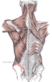 Muscles connecting the upper extremity to the vertebral column. Left iliac crest is labeled in red.
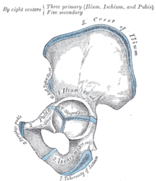 Plan of ossification of the hip bone.
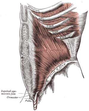 The Obliquus internus abdominis.
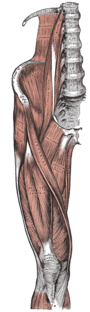 Muscles of the iliac and anterior femoral regions.
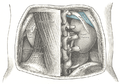 The relations of the kidneys from behind.
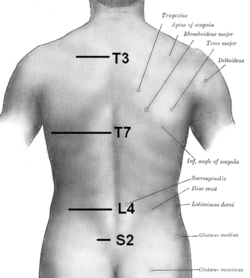 Iliac crest labeled at center right.
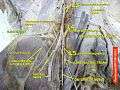 Iliac crest
See also
References
This article incorporates text in the public domain from page 234 of the 20th edition of Gray's Anatomy (1918)
- Platzer (2004), p 186
- Palastanga (2006), p 243
- Bogduk (2005), p 106
- Rose, Louis F.. Periodontics: Medicine, Surgery and Implants. Mosby, 072004. 26.2.2.3
- Bogduk, Nikolai; Endres, Stephen M. (2005). Clinical anatomy of the lumbar spine and sacrum (4th ed.). Elsevier Health Sciences. p. 106. ISBN 0-443-10119-1.
- Palastanga, Nigel; Field, Derek; Soames, Roger (2006). Anatomy and Human Movement: Structure and Function. Elsevier Health Sciences. ISBN 0-7506-8814-9.
- Platzer, Werner (2004). Color Atlas of Human Anatomy, Vol. 1: Locomotor System (5th ed.). Thieme. ISBN 3-13-533305-1.
External links
| Wikimedia Commons has media related to Iliac crest. |
- Anatomy figure: 13:02-01 at Human Anatomy Online, SUNY Downstate Medical Center - "Superficial muscles of the gluteal region and posterior thigh."
- Anatomy photo:35:os-0104 at the SUNY Downstate Medical Center - "Anterior Abdominal Wall: Osteology and Surface Anatomy "
- Atlas image: back_bone30 at the University of Michigan Health System - "The Back, Posterior View" (#4)
- Diagram at emedx.com
- Diagram at nih.gov
| Authority control |
|---|