Chin
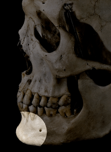
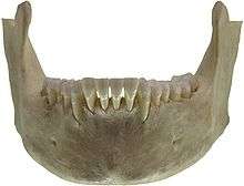
The human chin (also known as the mental protuberance, mental eminence, and rarely mental osseum, tuber symphyseos) refers to the forward pointed part of the anterior mandible (mental region) which is the area below the lower lip.
Evolution
The presence of a well-developed chin is considered to be one of the morphological characteristics of Homo sapiens that differentiates them from other human ancestors such as the closely related Neanderthals.[1][2] Early human ancestors have varied symphysial morphology, but none of them have a well-developed chin. Why modern humans have this particular feature and why it is not present in any fossil or living primate remain tantalizing questions. The origin of the chin is traditionally associated with the anterior–posterior breadth shortening of the dental arch or tooth row; however, its general mechanical or functional advantage during feeding, developmental origin, and link with human speech, physiology, and social influence are highly debated.
Functional perspectives
Robinson (1913)[3] suggests that the demand to resist masticatory stresses triggered bone thickening in the mental region of the mandible and ultimately formed a prominent chin. Moreover, Daegling (1993)[4] explains the chin as a functional adaptation to resist masticatory stress that causes vertical bending stresses in the coronal plane. Others have argued that the prominent chin is adapted to resisting wishboning forces,[5] dorso-ventral shear forces, and generally a mechanical advantage to resist lateral transverse bending and vertical bending in the coronal plane.[6] On the contrary, others[7] have suggested that the presence of the chin is not related to mastication. The presence of thick bone in the relatively small mandible may indicate better force resistance capacity. However, the question stands of whether the chin is an adaptive or nonadaptive structure.
Developmental perspectives
Recent works on the morphological changes of the mandible during development[8][9][10] have shown that the human chin, or at least the inverted-T shaped mental region, develops during the prenatal period, but the chin does not become prominent until the early postnatal period. This later modification happens by bone remodeling processes (bone resorption and bone deposition).[11] Coquerelle et al.[9][10] show that the anteriorly positioned cervical column of the spine and forward displacement of the hyoid bone limit the anterior–posterior breadth in the oral cavity for the tongue, laryngeal, and suprahyoid musculatures. Accordingly, this leads the upper parts of the mandible (alveolar process) to retract posteriorly, following the posterior movement of the upper tooth row, while the lower part of the symphysis remained protruded to create more space, thereby creating the inverted-T shaped mental relief during early ages and the prominent chin later. The alveolar region (upper or superior part of the symphysis) is sculpted by bone resorption, but the chin (lower or inferior part) is depository in its nature.[11] These coordinated bone growth and modeling processes mold the vertical symphysis present at birth into the prominent shape of the chin.
Recent research on the development of the chin[12] suggests that the evolution of this unique characteristic was formed not by mechanical forces such as chewing but by evolutionary adaptations involving reduction in size and change in shape of the face. Holton et al. claim that this adaptation occurred as the face became smaller compared to that of other ancient humans.
Other perspectives
Robert Franciscus takes a more anthropological viewpoint: he believes that the chin was formed as a consequence of the change in lifestyle humans underwent approximately 80,000 years ago. As humans began to turn their hunter-gatherer societies into agricultural societies where they increased their social networks, territorial disputes decreased because the new social structure promoted building alliances in order to exchange goods and belief systems. Franciscus believes that this change in the human environment reduced hormone levels, especially in men, resulting in the natural evolution of the chin.[13]
Overall, human beings are unique in the sense that they are the only species among primates who have chins. In the novel The Enduring Puzzle of the Human Chin, evolutionary anthropologists James Pampush and David Daegling discuss various theories that have been raised to solve the puzzle of the chin. They conclude that "each of the proposals we have discussed falter either empirically or theoretically; some fail, to a degree, on both accounts… This should serve as motivation, not discouragement, for researchers to continue investigating this modern human peculiarity… perhaps understanding the chin will reveal some unexpected insight into what it means to be human."[14]
Cleft chin
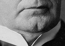
The terms cleft chin,[15] chin cleft,[15][16] dimple chin,[17][18] or chin dimple[15] refer to a dimple on the chin. It is a Y-shaped fissure on the chin with an underlying bony peculiarity.[19] Specifically, the chin fissure follows the fissure in the lower jaw bone that resulted from the incomplete fusion of the left and right halves of the jaw bone, or muscle, during the embryonal and fetal development. For other individuals, it can develop over time, often because one half of the jaw is longer than the other, leading to facial asymmetry.[15]
A cleft chin is an inherited trait in humans and can be influenced by many factors. The cleft chin is also a classic example of variable penetrance[20] with environmental factors or a modifier gene possibly affecting the phenotypical expression of the actual genotype. Cleft chins can be presented in a child when neither parent presents a cleft chin. Cleft chins are common among people originating from Europe, the Middle East and South Asia.[21]
In Persian literature, the chin dimple is considered a factor of beauty, and is metaphorically referred to as "the chin pit" or "the chin well": a well in which the poor lover is fallen and trapped.[22]
Double chin
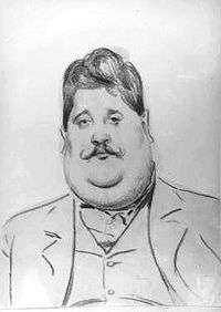
A double chin is a layer of subcutaneous fat around the neck that sags down and creates a wrinkle, creating the appearance of a second chin. This fat pad is occasionally surgically removed and the corresponding muscles under the jaw shortened.[23]
See also
- Chin augmentation (genioplasty)
- Masseter
- Mastication
- Otofacial syndrome
- Ptosis (chin)
References
- Stringer CB, Hublin JJ, Vandermeersch B (1984). The origin of anatomically modern humans in western Europe. In F. H. Smith & Spencer (Eds.), The origins of modern humans: a world survey of the fossil evidence. New York. pp. 51–135.
- Schwartz JH, Tattersall I (March 2000). "The human chin revisited: what is it and who has it?". Journal of Human Evolution. 38 (3): 367–409. doi:10.1006/jhev.1999.0339. PMID 10683306.
- Robinson L (1913). "The story of the chin". Knowledge. 36: 410–420.
- Daegling DJ (1993). "Functional morphology of the human chin". Evolutionary Anthropology: Issues, News, and Reviews. 1 (5): 170–177. doi:10.1002/evan.1360010506.
- DuBrul EL, Sicher H (1954). The Adaptive Chin. Springfield, IL: C.C. Thomas.
- Gröning F, Liu J, Fagan MJ, O'Higgins P (April 2011). "Why do humans have chins? Testing the mechanical significance of modern human symphyseal morphology with finite element analysis". American Journal of Physical Anthropology. 144 (4): 593–606. doi:10.1002/ajpa.21447. PMID 21404235.
- Ichim I, Swain M, Kieser JA (July 2006). "Mandibular biomechanics and development of the human chin". Journal of Dental Research. 85 (7): 638–42. doi:10.1177/154405910608500711. PMID 16798865.
- Coquerelle M, Bookstein FL, Braga J, Halazonetis DJ, Weber GW (November 2010). "Fetal and infant growth patterns of the mandibular symphysis in modern humans and chimpanzees (Pan troglodytes)". Journal of Anatomy. 217 (5): 507–20. doi:10.1111/j.1469-7580.2010.01287.x. PMC 3035859. PMID 20807267.
- Coquerelle M, Prados-Frutos JC, Rojo R, Mitteroecker P, Bastir M (2013-11-15). Frayer D (ed.). "Short faces, big tongues: developmental origin of the human chin". PLOS ONE. 8 (11): e81287. Bibcode:2013PLoSO...881287C. doi:10.1371/journal.pone.0081287. PMC 3829973. PMID 24260566.
- Coquerelle M, Prados-Frutos JC, Rojo R, Drake AG, Murillo-Gonzalez JA, Mitteroecker P (September 2017). "The Fetal Origin of the Human Chin". Evolutionary Biology. 44 (3): 295–311. doi:10.1007/s11692-017-9408-9. ISSN 0071-3260.
- Enlow DH (1990). Facial growth. 3rd edition. Philadelphia: Saunders.
- Holton NE, Bonner LL, Scott JE, Marshall SD, Franciscus RG, Southard TE (June 2015). "The ontogeny of the chin: an analysis of allometric and biomechanical scaling". Journal of Anatomy. 226 (6): 549–59. doi:10.1111/joa.12307. PMC 4450959. PMID 25865897.
- "Why we have chins: Our chin comes from evolution, not mechanical forces". ScienceDaily. April 13, 2015. Retrieved July 28, 2018.
- Gauger Ann (February 11, 2006). "On the origin of Chins". Evolution News. Retrieved July 28, 2018.
- Mammalian Phenotype Browser: Cleft chin
- Sharks of the world, Vol. 2, p. 143; by Leonard J. V. Compagno, Food and Agriculture Organization of the United Nations, 2001.
- Rob Roy, p. 229 (in 1872 edition, pub. Osgood); by Sir Walter Scott, 1817.
- "Myths of Human Genetics: Cleft Chin".
- Online Mendelian Inheritance in Man (OMIM) 119000
- Starr B. "Ask a Geneticist". Understanding Genetics. TheTech. Archived from the original on 2007-05-13. Retrieved 2007-07-01.
- Günther H. "Anomalien und Anomaliekomplexe in der Gegend des ersten Schlundbogens". Zeitschrift für menschliche Vererbungs- und Konstitutionslehre. 23: 43–52.; Lebow MR, Sawin PB (1941). "Inheritance of human facial features: a pedigree study involving length of face, prominent ears and chin cleft". Journal of Heredity. 32: 127–32. doi:10.1093/oxfordjournals.jhered.a105016.
- چاه زنخدان the chin well Archived 2014-08-08 at the Wayback Machine
- Larkin, Dimitrije E. Panfilov ; translated by Grahame (2005). Cosmetic surgery today. Stuttgart: Thieme Medical Publishers. p. 64. ISBN 1-58890-334-6.
| Wikimedia Commons has media related to Chins. |
| Authority control |
|---|