

Images of Influenza Viruses
 ShareCompartir
For images related to H7N9, go to Images of Avian Influenza A H7N9.
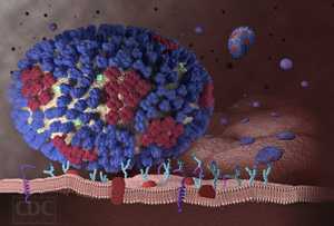
Understanding Influenza (Flu) Infection: An Influenza Virus Binds to a Respiratory Tract Cell
Description
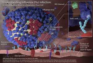
This image illustrates the very beginning stages of an influenza (flu) infection. Most experts think that influenza viruses spread mainly through small droplets containing influenza virus. These droplets are expelled into the air when people infected with the flu cough, sneeze or talk. Once in the air, these small infectious droplets can land in the mouths or noses of people who are nearby.This image shows what happens after these influenza viruses enter the human body. The viruses attach to cells within the nasal passages and throat (i.e., the respiratory tract). The influenza virus’s hemagglutinin (HA) surface proteins then bind to the sialic acid receptors on the surface of a human respiratory tract cell. The structure of the influenza virus’s HA surface proteins is designed to fit the sialic acid receptors of the human cell, like a key to a lock. Once the key enters the lock, the influenza virus is then able to enter and infect the cell. This marks the beginning of a flu infection.
Image with Labels and Full Text [JPG, 1.6 MB]
Image with Labels only [JPG, 1.4 MB]
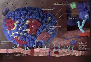
Image without Labels or Text [JPG, 1.3 MB]
![Download Image [JPG, 1.6 MB]](../flu/images/influenza-virus-fulltext.jpg)
![Download Image [JPG, 1.4 MB]](../flu/h/flu/images/influenza-virus-labels.jpg)
![Download Image [JPG, 1.3 MB]](../flu/images/influenza-virus-nolabels.jpg)
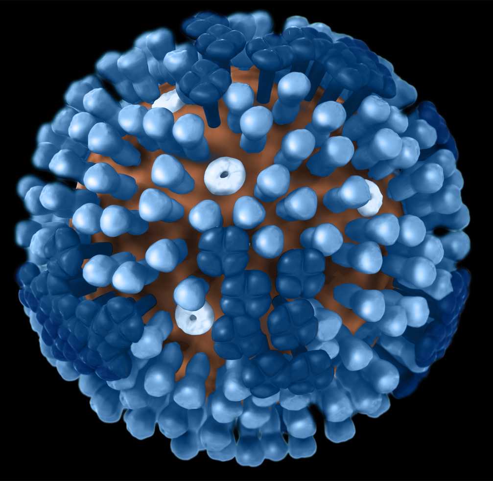
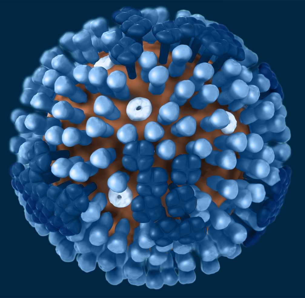
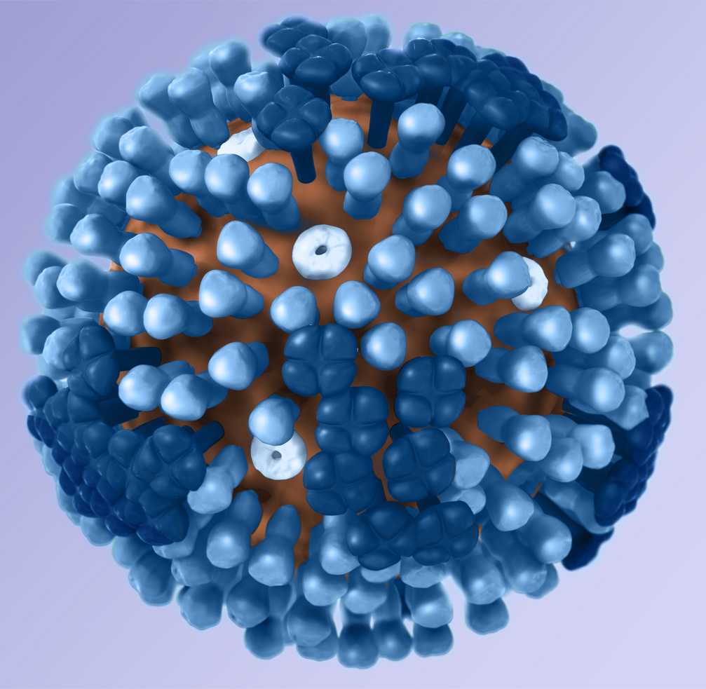
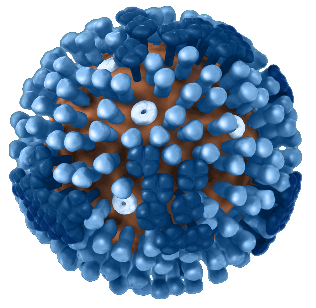
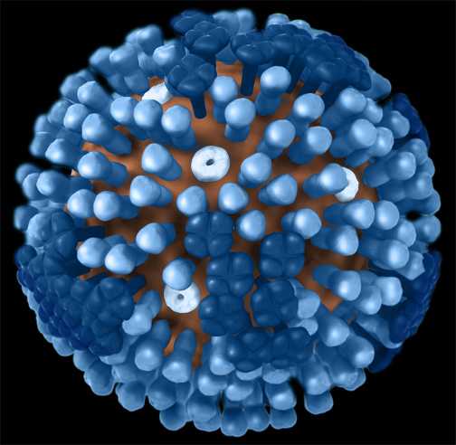
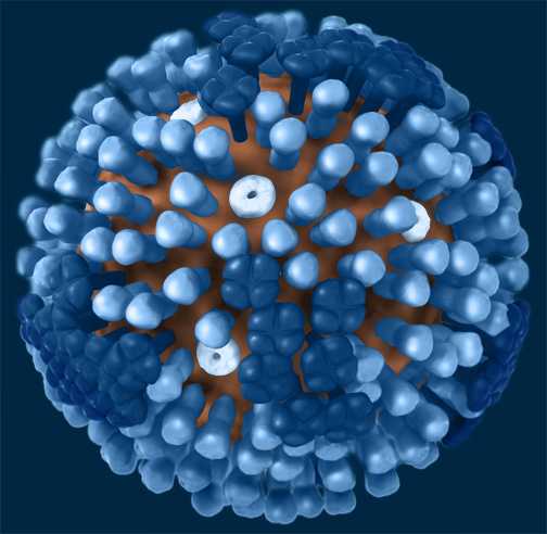
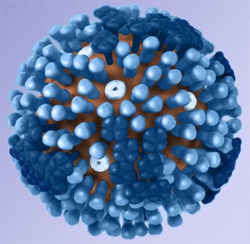
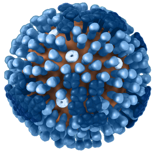
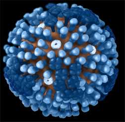
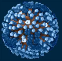
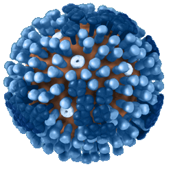
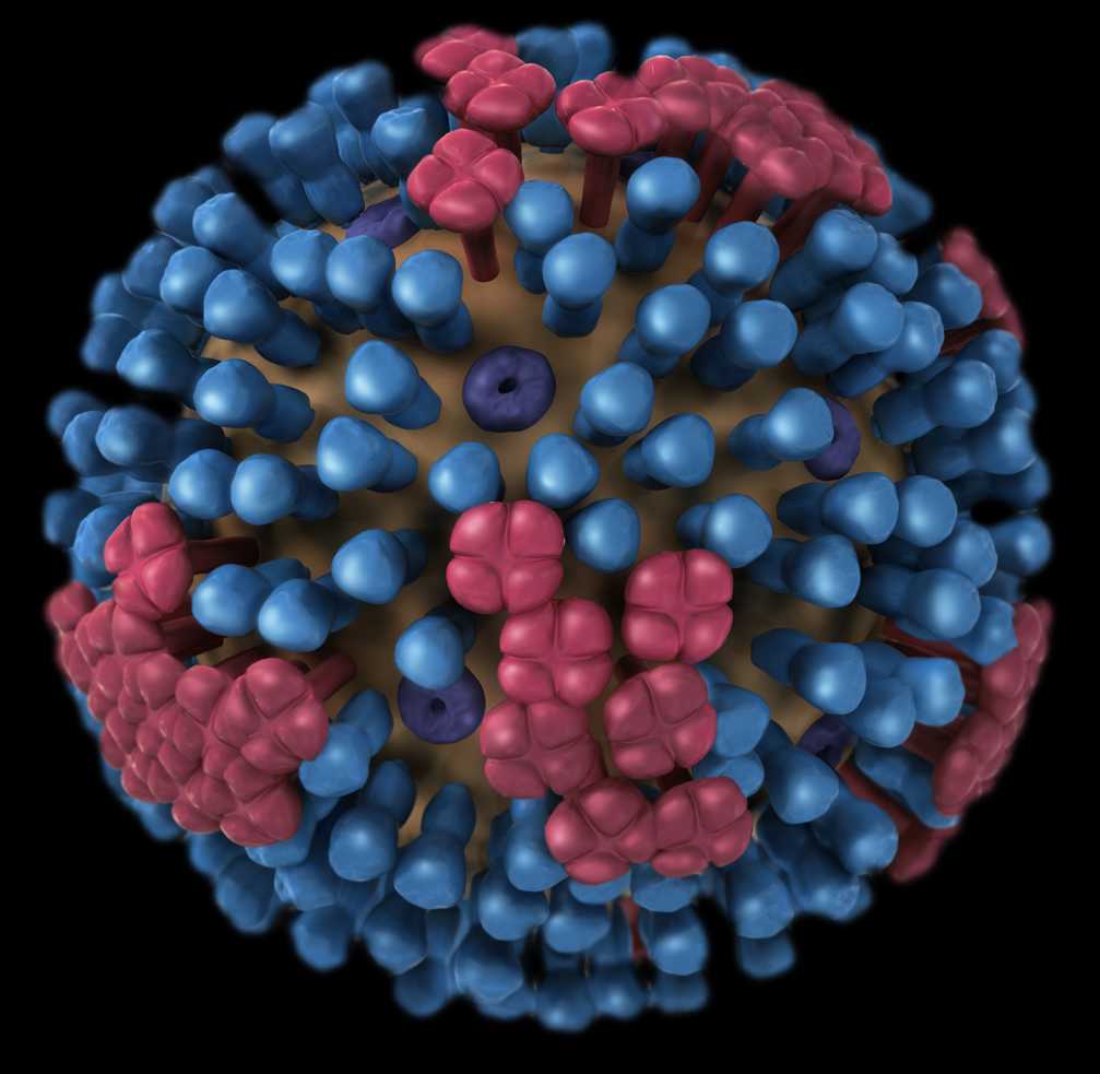
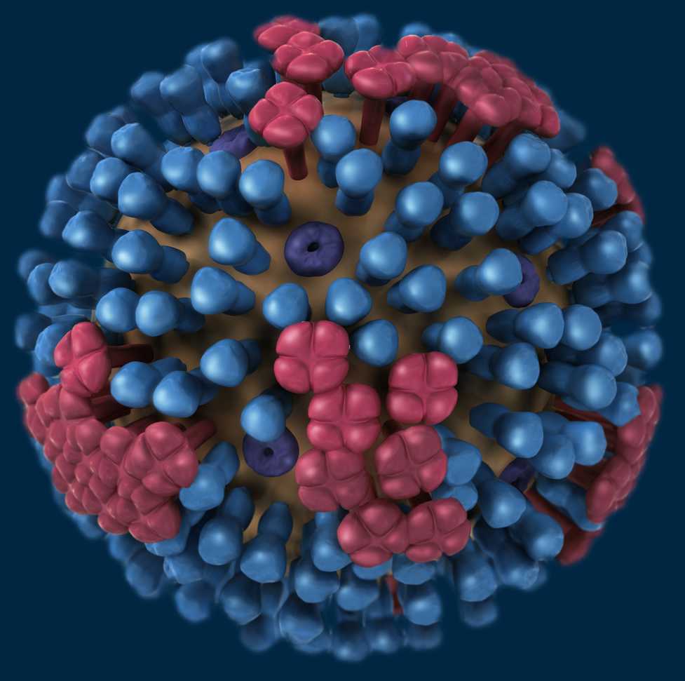
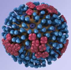
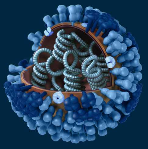
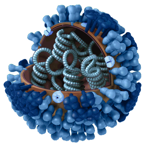
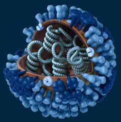
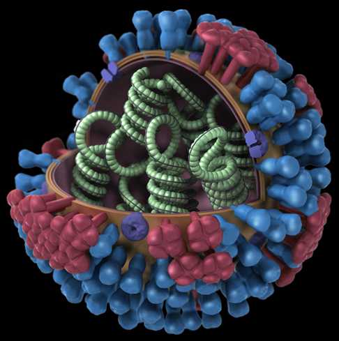
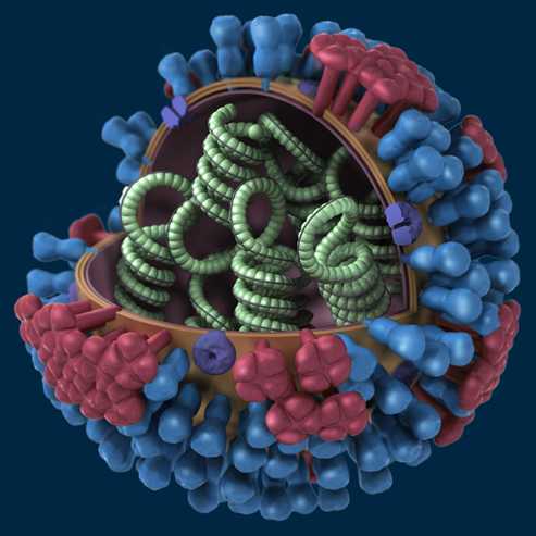
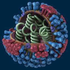
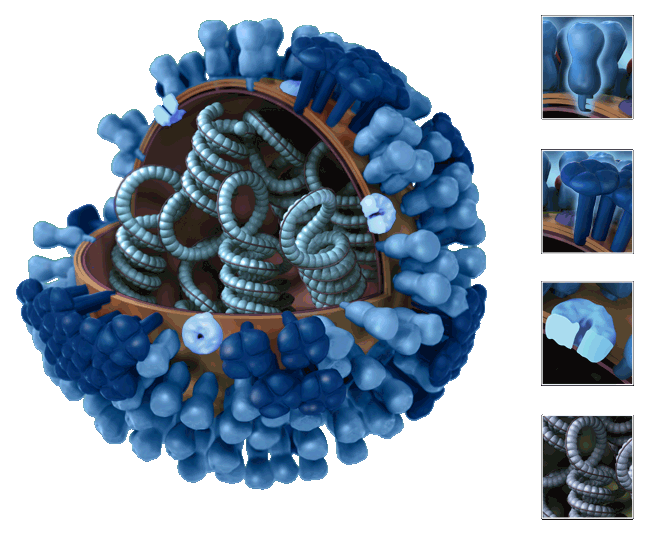
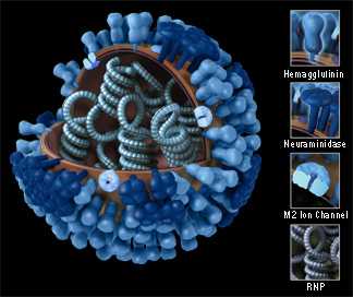
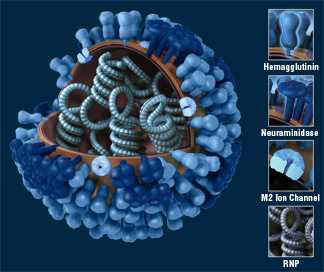
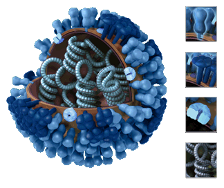
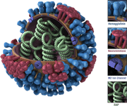
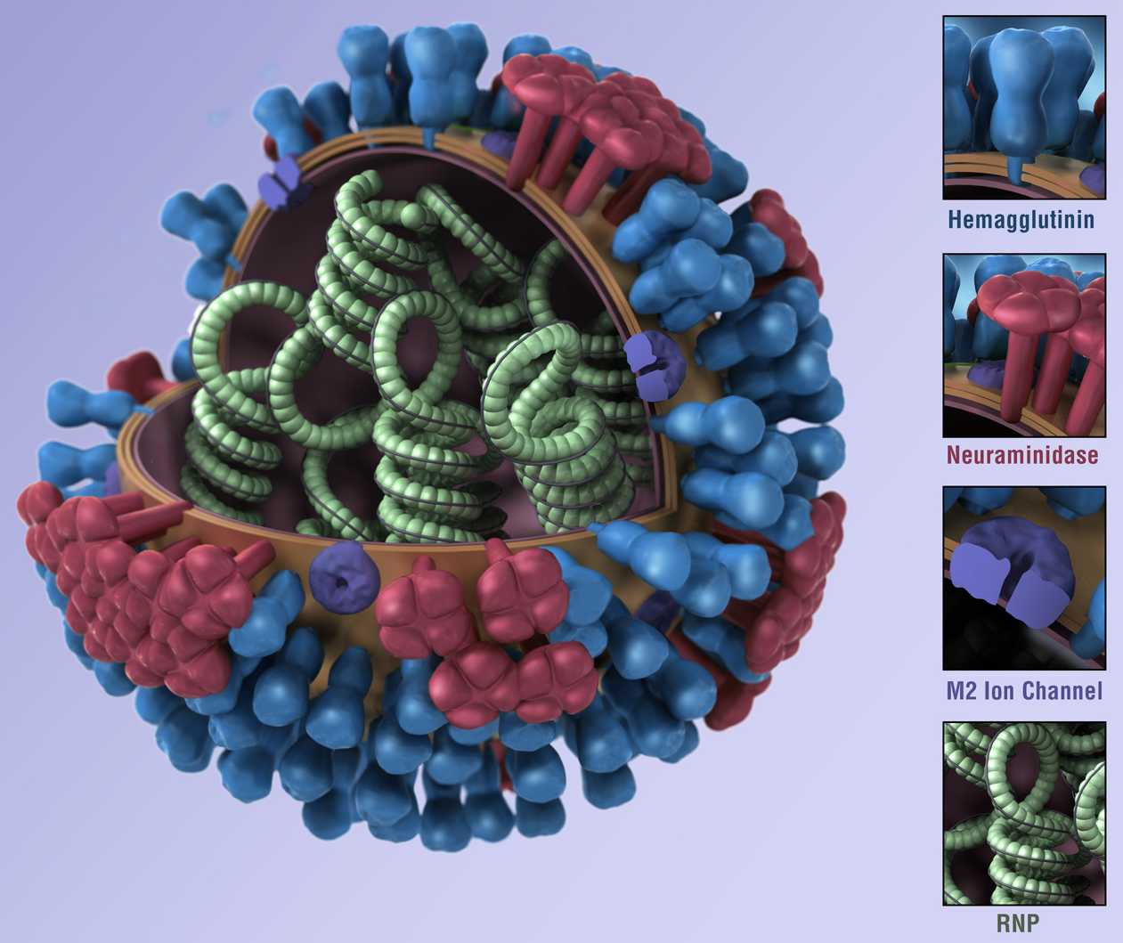
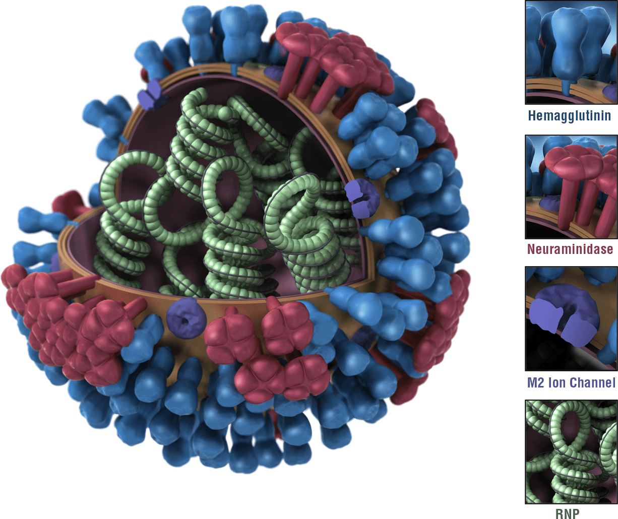
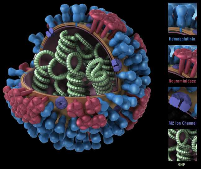
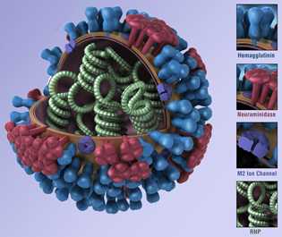
Graphical Representations of a Generic Influenza Virus
Description
These images provide a 3D graphical representation of the biology and structure of a generic influenza virus, and are not specific to the 2009 H1N1 virus. Available for download in 72 dpi.
3D View - Full - One Color
Available in these background colors — Posted March 26, 2010
|
Black | Blue | Purple | Transparent |
|---|---|---|---|---|
| Large | Large | Large | Large | |
| Medium | Medium | Medium | Medium | |
| Small | Small | Small | Small |
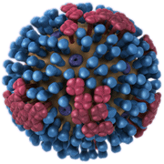
3D View - Full - Multiple Colors
Available in these background colors — Posted November 25, 2009
|
Black | Blue | Purple | Transparent |
|---|---|---|---|---|
| Large | Large | Large | Large | |
| Medium | Medium | Medium | Medium | |
| Small | Small | Small | Small |
3D View - Sliced - One Color
Available in these background colors — Posted March 26, 2010
|
Black | Blue | Transparent |
|---|---|---|---|
| Large | Large | Large | |
| Medium | Medium | Medium | |
| Small | Small | Small |
3D View - Sliced - Multiple Colors
Available in these background colors — Posted November 25, 2009
|
Black | Blue | Transparent |
|---|---|---|---|
| Large | Large | Large | |
| Medium | Medium | Medium | |
| Small | Small | Small |
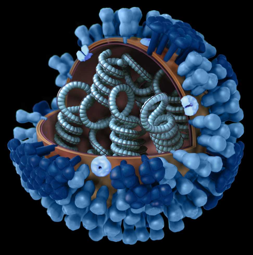
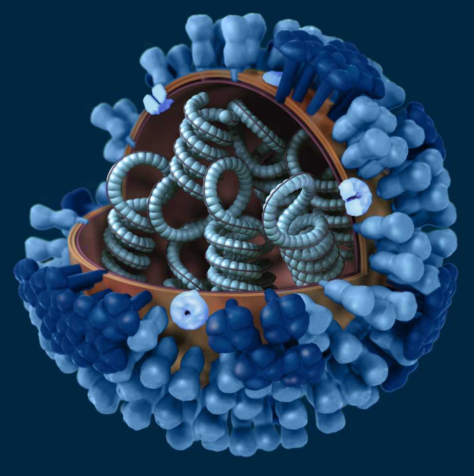
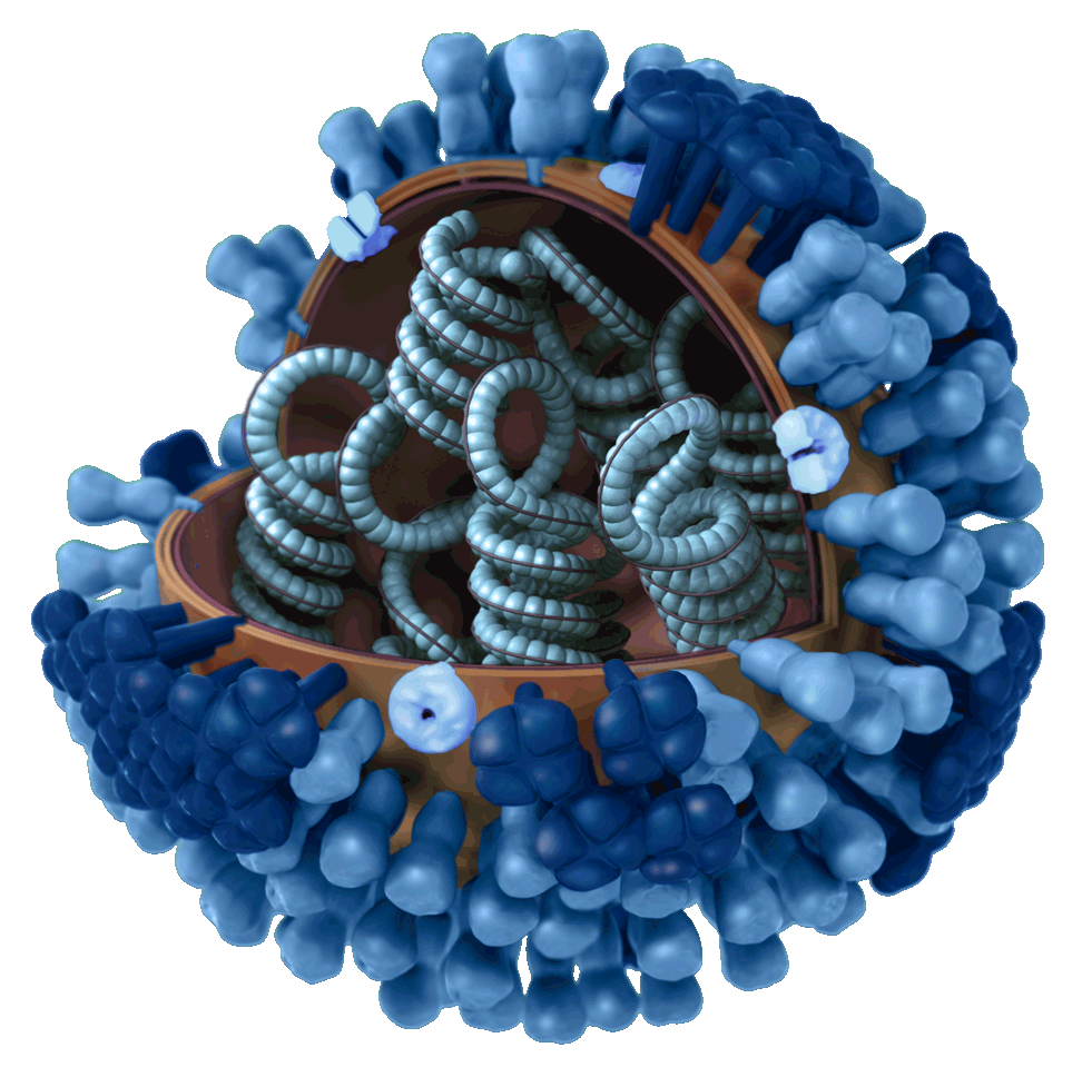
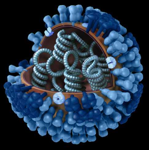
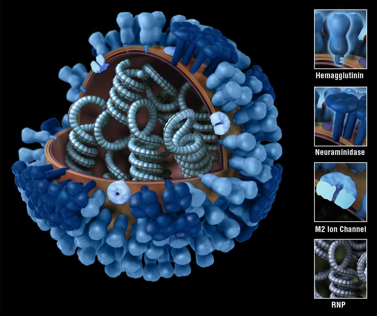
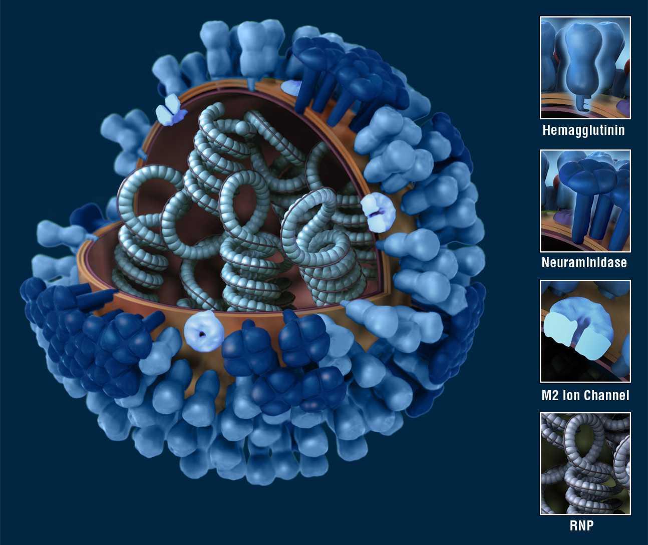
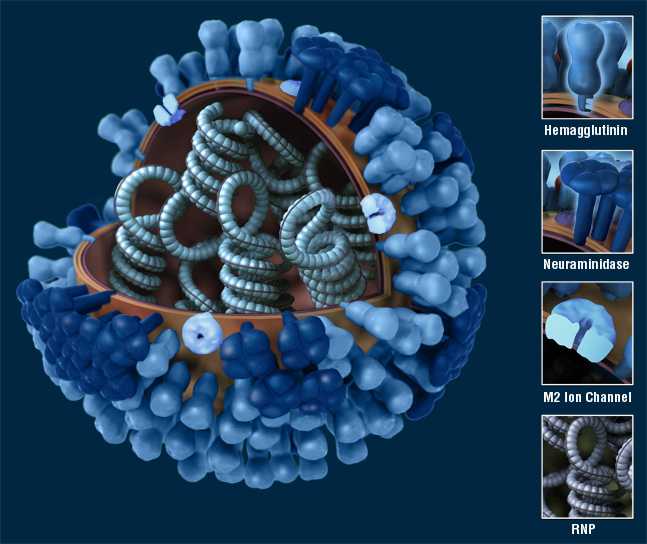
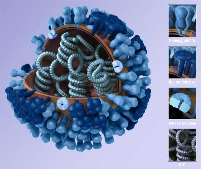
3D View - Sliced with Key - One Color
Available in these background colors — Posted March 26, 2010
|
Black | Blue | Purple | Transparent |
|---|---|---|---|---|
| Large | Large | Large | Large | |
| Medium | Medium | Medium | Medium | |
| Small | Small | Small | Small |
3D View - Sliced with Key - Multiple Colors
Available in these background colors — Posted November 25, 2009
|
Black | Blue | Purple | Transparent |
|---|---|---|---|---|
| Large | Large | Large | Large | |
| Medium | Medium | Medium | Medium | |
| Small | Small | Small | Small |
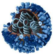
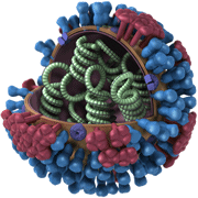
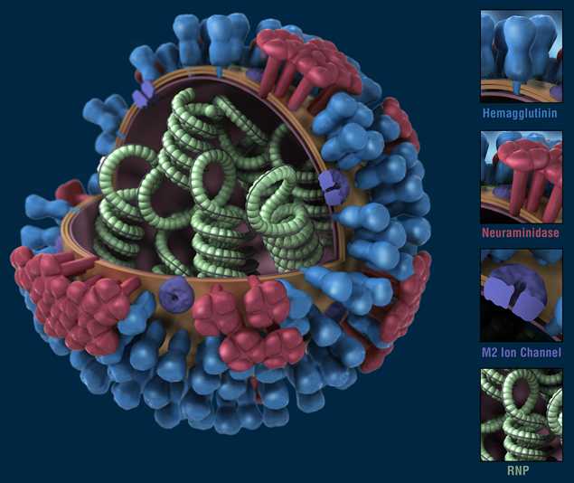
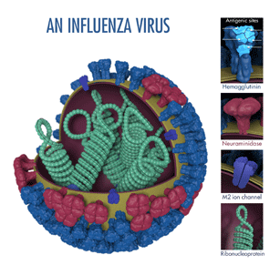
3D View - Sliced with Key - Antigenic Sites
Available on a white background — Posted May 19, 2014
|
White |
|---|---|
| Large | |
| Medium | |
| Small |
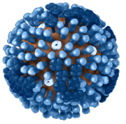
The above image shows the different features of an influenza virus, including the surface proteins hemagglutinin (HA) and neuraminidase (NA). Following influenza infection or receipt of the influenza vaccine, the body’s immune system develops antibodies that recognize and bind to “antigenic sites,” which are regions found on an influenza virus’ surface proteins. By binding to these antigenic sites, antibodies neutralize flu viruses, which prevents them from causing further infection.
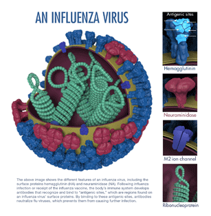
3D View - Sliced with Key - Antigenic Sites with Caption
Available on a white background with captions — Posted June 4, 2014
|
White |
|---|---|
| Large | |
| Medium | |
| Small |
The above image shows the different features of an influenza virus, including the surface proteins hemagglutinin (HA) and neuraminidase (NA). Following influenza infection or receipt of the influenza vaccine, the body’s immune system develops antibodies that recognize and bind to “antigenic sites,” which are regions found on an influenza virus’ surface proteins. By binding to these antigenic sites, antibodies neutralize flu viruses, which prevents them from causing further infection.
- Page last reviewed: June 4, 2014
- Page last updated: August 15, 2014
- Content source:
- Centers for Disease Control and Prevention, National Center for Immunization and Respiratory Diseases (NCIRD)
- Page maintained by: Office of the Associate Director for Communication, Digital Media Branch, Division of Public Affairs