Tarsal tunnel
The tarsal tunnel is found along the inner leg posterior to the medial malleolus.
| Tarsal tunnel | |
|---|---|
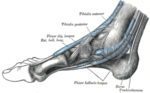 The mucous sheaths of the tendons around the ankle. Medial aspect. | |
| Details | |
| Identifiers | |
| Latin | Canalis tarsi |
| Anatomical terminology | |
Structure
The tarsal tunnel is made up of bone on the inside and the flexor retinaculum on the outside.
Function
Contents
The tibial nerve, posterior tibial artery, posterior tibial vein, and flexor tendons travel in a bundle along this pathway through the tarsal tunnel, in the following order from antero-medial to postero-lateral:
- Tibialis posterior tendon
- Flexor digitorum longus tendon
- Posterior tibial artery
- Posterior tibial vein
- Tibial nerve
- Flexor hallucis longus tendon
In the tunnel, the nerve splits into three different paths. One nerve (calcaneal) continues to the heel, the other two (medial plantar nerve and lateral plantar nerve) continue on to the bottom of the foot.
Clinical significance
Tarsal tunnel syndrome
Tarsal tunnel syndrome is the most commonly reported nerve entrapment of the ankle and is analogous to the carpal tunnel of the wrist. People with tarsal tunnel syndrome have pain in the plantar aspect of the foot mostly at night. Weight bearing increases pain and weakness is found on intrinsic foot muscles with positive Tinel sign at the tunnel. There is no tenderness present on the plantar foot, though this is typically the primary site of complaint.
Additional images
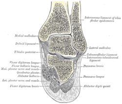 Coronal section through right talocrural and talocalcaneal joints.
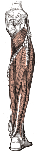 Muscles of the back of the leg. Deep layer.
 The popliteal, posterior tibial, and peroneal arteries.
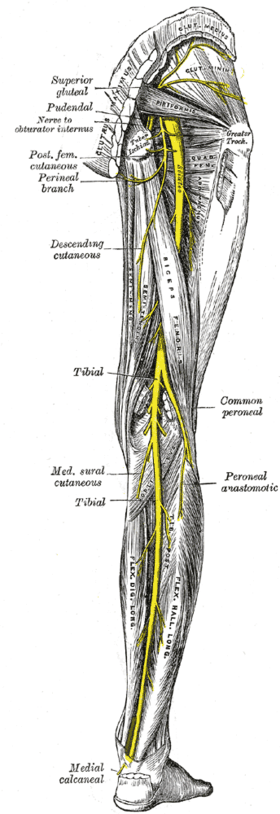 Nerves of the right lower extremity Posterior view.
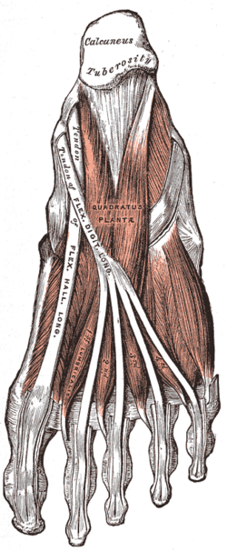 Muscles of the sole of the foot. Second layer.
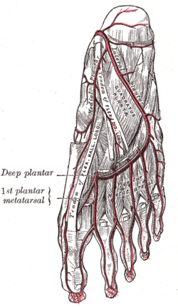 The plantar arteries. Deep view.
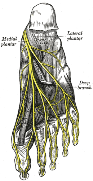 The plantar nerves.
See also
References
External links
| Wikimedia Commons has media related to Tarsal tunnel. |