Plantaris muscle
The plantaris is one of the superficial muscles of the superficial posterior compartment of the leg, one of the fascial compartments of the leg.
| Plantaris muscle | |
|---|---|
 Animation | |
 The plantaris is visible under the gastrocnemius. | |
| Details | |
| Origin | Lateral supracondylar ridge of femur above lateral head of gastrocnemius |
| Insertion | Tendo calcaneus (medial side, deep to gastrocnemius tendon) |
| Artery | sural arteries |
| Nerve | tibial nerve from anterior rami of S1-S2 |
| Actions | Plantar flexes foot and flexes knee |
| Antagonist | Tibialis anterior muscle |
| Identifiers | |
| Latin | musculus plantaris |
| TA | A04.7.02.049 |
| FMA | 22543 |
| Anatomical terms of muscle | |
It is composed of a thin muscle belly and a long thin tendon. While not as thick as the achilles tendon, the plantaris tendon (which tends to be between 30–45 centimetres (12–18 in) in length) is the longest tendon in the human body. Not including the tendon, the plantaris muscle is approximately 5–10 centimetres (2.0–3.9 in) long and is absent in 8-12% of the population. It is one of the plantar flexors in the posterior compartment of the leg, along with the gastrocnemius and soleus muscles. The plantaris is considered an unimportant muscle and mainly acts with the gastrocnemius.
Structure
The plantaris muscle arises from the inferior part of the lateral supracondylar ridge of the femur at a position slightly superior to the origin of the lateral head of gastrocnemius. It passes posterior to the knee joint in an inferomedial direction and becomes tendinous distally to insert into the Achilles tendon. It occasionally separately inserts into the medial side of the calcaneus.
Innervation
The plantaris muscle is innervated by the tibial nerve, a branch of the sciatic nerve in the sacral plexus. Signaling for contraction begins in the frontal lobe of the brain with the pre-central gyrus (primary motor cortex). Upper motor neurons are stimulated and send a signal through the internal capsule and down the corticospinal tract. Decussation of the lateral corticospinal tract occurs in the medullary pyramids, then the fibers continue down the contralateral side of the spinal cord. Upper motor neurons synapse with lower motor neurons at the anterior horn of the spinal cord in the sacral plexus (formed from the anterior rami of spinal nerves L4, L5, S1–4). The lower motor neuron fibers continue down the sciatic nerve and then diverge into the tibial and common fibular nerves. The tibial nerve runs medially at the knee joint. When the tibial nerve receives an action potential, the plantaris muscle contracts, providing weak plantar flexion of the foot and weak flexion of the knee.[1]
Variation
The muscle may arise from the oblique popliteal ligament. Interdigitations with the lateral head of the gastrocnemius and a fibrous extension of the muscle to the patella are not unusual.[2]
Function
The plantaris acts to weakly plantar flex the ankle joint and flex the knee joint.
The plantaris muscle may also provide proprioceptive feedback information to the central nervous system regarding the position of the foot. The unusually high density of proprioceptive receptor end organs supports this notion.[3]
Its motor function is so minimal that its long tendon can readily be harvested for reconstruction elsewhere with little functional deficit. Often mistaken for a nerve by new medical students (and thus called the "freshman nerve"), the muscle was useful to other primates for grasping with their feet.[4]
Clinical significance
A common injury that is normally attributed to the plantaris muscle is a condition called tennis leg. Although pain in the calf can be attributed to a rupture of the plantaris muscle, recent ultrasound research has shown that tennis leg more commonly arises from tears in the musculotendinous junction of the medial gastrocnemius. In one clinical study, 94 out of 141 patients (66.7%) diagnosed with tennis leg were found with a partial rupture of the gastrocnemius muscle, while rupture of the plantaris tendon was only seen in 2 patients (1.4%).[5]
Injury may occur from running, jumping, or pushing off one leg in sports such as tennis, basketball and soccer, which require quick foot movement in a certain direction. Isolated plantaris muscle strains are rare, and ruptures normally occur in conjunction with injury to other muscles in the posterior compartment of the lower leg.[6] Symptoms of a plantaris muscle rupture may include an audible popping sound in the area during physical activity, swelling, pain in the back of the lower leg, and persistent soreness. Ankle flexion may also be painful.[7]
See also
Additional images
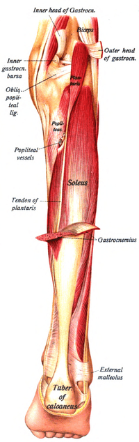
 Tendon of Plantaris is labeled under the gastrocnemius (left), and folded away (right).
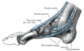 The synovial sheaths of the tendons around the ankle. Medial aspect. (Tendon of Plantaris labeled at bottom right.)
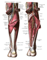
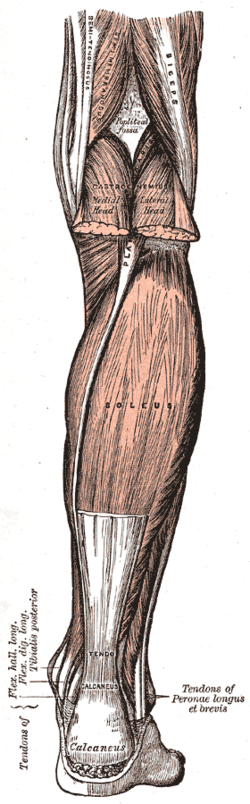 Muscles of the back of the leg. Superficial layer.
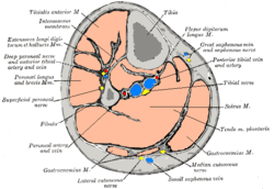 Cross-section through middle of leg.
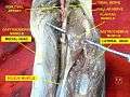 Plantaris muscle
References
- Saladin, Kenneth S. Anatomy and Physiology The Unity of Form and Function. 6th ed. New York: McGraw-Hill Science Engineering, 2009. Print.
- Freeman, A. J.; Jacobson, N. A.; Fogg, Q. A. (2008). "Anatomical variations of the plantaris muscle and a potential role in patellofemoral pain syndrome". Clinical Anatomy. 21 (2): 178–81. doi:10.1002/ca.20594. PMID 18266282.
- Moore, Keith L; & Dalley Arthur R (2008). Clinically Oriented Anatomy (6th ed.). Lippincott Williams and Wilkins. ISBN 978-1-60547-652-0
- Andor, W.J.M., Glaudemans, Rudi A.J.O. Dierckx, Jan L.M.A. Gielen, Johannes (Hans) Zwerver (2015). Nuclear Medicine and Radiologic Imaging in Sports Injuries. Springer. p. 762
- Delgado, Gonzalo J.; Chung, Christine B.; Lektrakul, Nitaya; Azocar, Patricio; Botte, Michael J.; Coria, Daniel; Bosch, Enrique; Resnick, Donald (2002). "Tennis Leg: Clinical US Study of 141 Patients and Anatomic Investigation of Four Cadavers with MR Imaging and US1". Radiology. 224 (1): 112–9. doi:10.1148/radiol.2241011067. PMID 12091669.
- Spina, A. A. (2007). "The plantaris muscle: Anatomy, injury, imaging, and treatment". The Journal of the Canadian Chiropractic Association. 51 (3): 158–65. PMC 1978447. PMID 17885678.
- http://www.livestrong.com/article/510804-running-injuries-to-the-plantaris-soleus-muscles/%5B%5D
External links
| Wikimedia Commons has media related to Plantaris muscle. |
- Anatomy photo:15:st-0412 at the SUNY Downstate Medical Center
- PTCentral
| Authority control |
|---|