Femoral sheath
The femoral sheath (crural sheath) is formed by a prolongation downward, behind the inguinal ligament, of the abdominal fascia, the transverse fascia being continued down in front of the femoral vessels and the iliac fascia behind them. The femoral sheath is contained within the femoral triangle.
| Femoral sheath | |
|---|---|
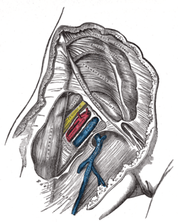 Femoral sheath laid open to show its three compartments. | |
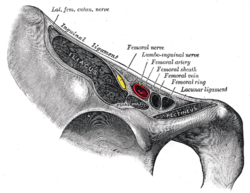 Structures passing behind the inguinal ligament. | |
| Anatomical terminology |
The sheath assumes the form of a short funnel, the wide end of which is directed upward, while the lower, narrow end fuses with the fascial investment of the vessels, about 4 cm. below the inguinal ligament.
It is strengthened in front by a band termed the iliopubic tract.
The lateral wall of the sheath is vertical and is perforated by the lumboinguinal nerve; the medial wall is directed obliquely downward and lateralward, and is pierced by the great saphenous vein and by some lymphatic vessels.
The sheath is divided by two vertical partitions which stretch between its anterior and posterior walls.
The lateral compartment contains the femoral artery and femoral branch of genitofemoral nerve, and the intermediate the femoral vein, while the medial and smallest compartment is named the femoral canal, and contains some lymphatic vessels and a lymph gland embedded in a small amount of areolar tissue.
The femoral canal is conical and measures about 1.25 cm. in length. Its base, directed upward and named the femoral ring, is oval in form, its long diameter being directed transversely and measuring about 1.25 cm.
The spermatic cord in the male and the round ligament of the uterus in the female lie immediately above the anterior margin of the ring, while the inferior epigastric vessels are close to its upper and lateral angle.
The femoral ring is closed by a somewhat condensed portion of the extraperitoneal fatty tissue, named the septum femorale (crural septum), the abdominal surface of which supports a small lymph gland and is covered by the parietal peritoneum.
The septum femorale is pierced by numerous lymphatic vessels passing from the deep inguinal to the external iliac lymph glands, and the parietal peritoneum immediately above it presents a slight depression named the femoral fossa.
Additional Images
 Femoral sheath
References
This article incorporates text in the public domain from page 625 of the 20th edition of Gray's Anatomy (1918)
External links
- Photo and overview at gla.ac.uk
- antthigh at The Anatomy Lesson by Wesley Norman (Georgetown University)
- Diagram at washington.edu