Adductor muscles of the hip
The adductor muscles of the hip are a group of muscles mostly used for bringing the thighs together (called adduction).
| Adductor muscles of the hip | |
|---|---|
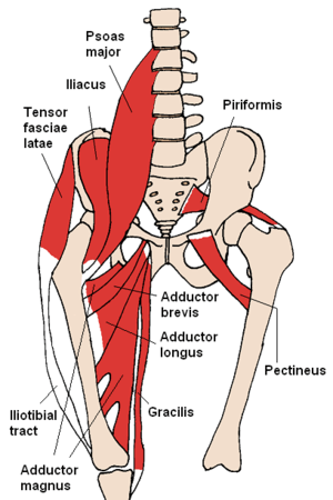 The adductors and nearby muscles | |
| Details | |
| Origin | Pubis |
| Insertion | Femur and tibia |
| Nerve | Obturator nerve |
| Actions | Adduction of hip |
| Anatomical terms of muscle | |
Structure
The adductor group is made up of:
- Adductor brevis
- Adductor longus
- Adductor magnus
- Adductor minimus This is often considered to be a part of adductor magnus.
- pectineus
- gracilis
- Obturator externus[1] and are also part of the medial compartment of thigh
The adductors originate on the pubis and ischium bones and insert mainly on the medial posterior surface of the femur.
| Muscle | Origin | Insertion | innervation[2] |
| Adductor brevis | Inferior pubic ramus | Medial ridge of linea aspera | Obturator nerve (L2-L4) |
| Adductor longus | Front side of the pubic bone under the pubic tubercle | Medial ridge of linea aspera | Obturator nerve (L2-L4) |
| Adductor magnus | Inferior pubic ramus and ischial tuberosity | Medial ridge of linea aspera and the adductor tubercle | Obturator nerve and tibial nerve (L2-L5) |
| Adductor minimus | Inferior pubic ramus | Medial ridge of linea aspera | Obturator nerve (L2)[3] |
| Pectineus | Pectineal line (pubis) | Pectineal line | Femoral nerve and sometimes the obturator nerve (L2-L4) |
| Gracilis | Inferior pubic ramus | Pes anserinus on the tibia | Obturator nerve (L2-L3) |
| Obturator externus | Lateral surface of obturator membrane and the ischiopubic ramus | Trochanteric fossa | Posterior branch of obturator nerve (L5-S2) |
Nerve supply
The pectineus is the only adductor muscle that is innervated by the femoral nerve. The other adductor muscles are innervated by the obturator nerve[1] with the exception of a small part of the adductor magnus which is innervated by the tibial nerve.[4]
Variation
In 33% of people a supernumerary muscle is found between the adductor brevis and adductor minimus. When present, this muscle originates from the upper part of the inferior ramus of the pubis from where it runs downwards and laterally. In half of cases, it inserts into the anterior surface of the insertion aponeurosis of the adductor minimus. In the remaining cases, it is either inserted into the upper part of the pectineal line or the posterior part of the lesser trochanter. While similar to its neighbouring adductors, it is formed by separation from the superficial layer of the obturator externus, and is thus not ontogenetically related to the adductors.[5]
Clinical significance
Tenotomy
So-called adductor tenotomy (cutting the origin tendons of the adductor muscles of the thigh) and obturator neurectomy (cutting the anterior branch of the obturator nerve) is sometimes performed on children with cerebral palsy. These children often have hypertonia of the adductor muscles, making abduction difficult, obstructing normal hip development, and putting them at risk of hip luxation.
References
- Platzer, Werner (2004), " Color Atlas of Human Anatomy, Vol. 1, Locomotor System, Thieme, 5th ed, p 240
- Bojsen-Møller, Finn; Simonsen, Erik B.; Tranum-Jensen, Jørgen (2001). Bevægeapparatets anatomi [Anatomy of the Locomotive Apparatus] (in Danish) (12th ed.). pp. 364–367. ISBN 978-87-628-0307-7.
- "Adductor minimus". AnatomyExpert. Archived from the original on April 25, 2013. Retrieved April 30, 2013.
- Bojsen-Møller, Finn; Simonsen, Erik B.; Tranum-Jensen, Jørgen (2001). Bevægeapparatets anatomi [Anatomy of the Locomotive Apparatus] (in Danish) (12th ed.). p. 266. ISBN 978-87-628-0307-7.
- Nakamura E, Masumi S, Miura M, Kato S, Miyauchi R (August 1992). "A supernumerary muscle between the adductors brevis and minimus in humans". Okajimas Folia Anat Jpn. Okajimas Folia Anat Jpn. 1992 Aug;69(2-3):89-98. 69 (2–3): 89–98. PMID 1436954.