Tensor fasciae latae muscle
The tensor fasciae latae (or tensor fasciæ latæ or, formerly, tensor vaginae femoris) is a muscle of the thigh. Together with the gluteus maximus, it acts on the iliotibial band and is continuous with the iliotibial tract, which attaches to the tibia. The muscle assists in keeping the balance of the pelvis while standing, walking, or running.
| Tensor fasciae latae | |
|---|---|
 The tensor fasciae latae and nearby muscles | |
| Details | |
| Pronunciation | /ˈtɛnsər ˈfæʃii ˈleɪti/ |
| Origin | Anterior superior iliac spine |
| Insertion | Iliotibial tract (via greater trochanter) |
| Artery | Primarily lateral circumflex femoral artery, superior gluteal artery |
| Nerve | Superior gluteal nerve (L4, L5, S1) |
| Actions | Hip - flexion, medial rotation, abduction, knee - lateral rotation, Torso - stabilization |
| Identifiers | |
| Latin | Musculus tensor fasciae latae |
| TA | A04.7.02.010 |
| FMA | 22423 |
| Anatomical terms of muscle | |
Structure
It arises from the anterior part of the outer lip of the iliac crest; from the outer surface of the anterior superior iliac spine, and part of the outer border of the notch below it, between the gluteus medius and sartorius; and from the deep surface of the fascia lata.
It is inserted between the two layers of the iliotibial tract of the fascia lata about the junction of the middle and upper thirds of the thigh. The tensor fasciae latae tautens the iliotibial tract and braces the knee, especially when the opposite foot is lifted.[1] The terminal insertion point lies on the lateral condyle of the tibia.[2]
Nerve supply
Tensor fasciae latae is innervated by the superior gluteal nerve, L5 and S1. At its origins of the anterior rami of L4, L5, and S1 nerves, the superior gluteal nerve exits the pelvis via greater sciatic foramen superior to the piriformis. The nerve also courses between the gluteus medius and minimus. The superior gluteal artery also supplies the tensor fasciae latae.[1] The superior gluteal nerve arises from the sacral plexus and only has muscular innervation associated with it. There is no cutaneous innervation for sensation that stems from the superior gluteal nerve.[2]
Function
The tensor fasciae latae is a tensor of the fascia lata; continuing its action, the oblique direction of its fibers enables it to stabilize the hip in extension (assists gluteus maximus during hip extension). The fascia lata is a fibrous sheath that encircles the thigh like a subcutaneous stocking and tightly binds its muscles. On the lateral surface, it combines with the tendons of the gluteus maximus and tensor fasciae latae to form the iliotibial tract, which extends from the iliac crest to the lateral condyle of the tibia.[1]
In the erect posture, acting from below, it will serve to steady the pelvis upon the head of the femur; and by means of the iliotibial tract it steadies the condyles of the femur on the articular surfaces of the tibia, and assists the gluteus maximus in supporting the knee in a position of extension.
The basic functional movement of tensor fasciae latae is walking. The tensor fasciae latae is heavily utilized in horse riding, hurdling and water skiing. Some problems that arise when this muscle is tight or shortened are pelvic imbalances that lead to pain in hips, as well as pain in the lower back and lateral area of knees.[3]
Because of its insertion point on the lateral condyle of the tibia, it also aids in the lateral rotation of the tibia. This lateral rotation may be initiated in conjunction with hip abduction and medial rotation of the femur while kicking a soccer ball. The tensor fasciae latae works in synergy with the gluteus medius and gluteus minimus muscles to abduct and medially rotate the femur.
The TFL is a hip abductor muscle. To stretch the tensor fasciae latae, the knee may be brought medially across the body (adducted). If one leans against a wall with crossed legs (externally/laterally rotated hips) and pushes the pelvis away from the wall (leaning the upper body towards it) sidebending the lumbar spine (i.e.: curving the spine to the side) should be avoided as it stretches the lumbar region rather than the tensor fasciae latae and other muscles which cross the hip rather than the spine.
Clinical significance
Because it is used for so many movements and is in a shortened position when seated, the TFL becomes tight easily. TFL stretches lengthen this important muscle.[4]
Etymology
"Tensor fasciae latae" translates from Latin to English as "stretcher of the side band". "Tensor" is an agent noun that comes from the past participle stem "tens-" of the Latin verb "tendere", meaning "to stretch".[6] "Fasciae" is the Latin term for "of the band" and is in the singular genitive case. "Latae" is the respective singular, genitive, feminine form of the Latin adjective "latus" meaning "side".[7][8]
Additional images
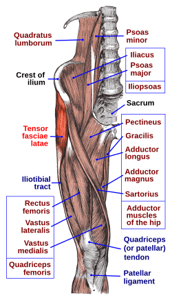
 Muscles of the pelvis and upper leg, showing the tensor fasciae latae at the middle left.
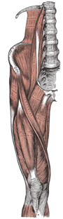
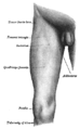 Front of the right thigh, with the position of the tensor fasciae latae indicated.
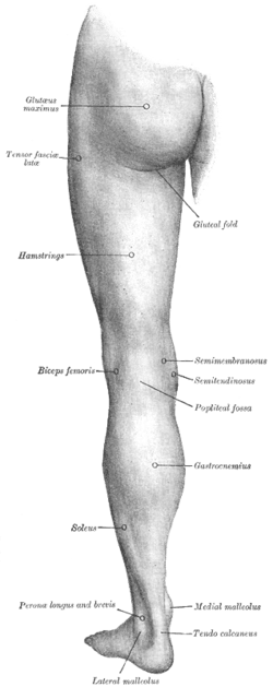 Back of the left thigh, with the position of the tensor fasciae latae indicated.
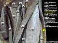 Tensor fasciae latae muscle
 Tensor fasciae latae muscle
References
This article incorporates text in the public domain from page 476 of the 20th edition of Gray's Anatomy (1918)
- Saladin, Kenneth. Anatomy and Physiology. 6th ed. Mc-Graw Hill. 2010.
- Saladin, Kenneth. Anatomy and Physiology. 5th ed. Mc-Graw Hill. 2010.
- Jarmey, Chris. The Concise Book of Muscles. North Atlantic Books: Berkeley, CA, 2003. 2nd Ed.
- Joe Miller (2015-05-13). "Tensor Fasciae Latae Muscle Stretches". Livestrong.com. Retrieved 2015-05-31.
- Case Studies:Jonathan M. Cooperman, Journal of Orthopaedic & Sports Physical Therapy 1984 5:4, 201-203, DOI: 10.2519/jospt.1984.5.4.201
- "Online Etymology Dictionary". Etymonline.com. Retrieved 2015-05-31.
- "Tensor fasciae latae - Structure Detail". Anatomyexpert.com. Retrieved 2015-05-31.
- "Latin Noun Declensions". Sacredbible.org. Retrieved 2015-05-31.
External links
| Wikimedia Commons has media related to Tensor fasciae latae muscle. |
- Cross section image: pelvis/pelvis-e12-15—Plastination Laboratory at the Medical University of Vienna
- Muscles/TensorFasciaeLatae at exrx.net
- Coachr
| Authority control |
|---|