Semimembranosus muscle
The semimembranosus (/ˌsɛmiˌmɛmbrəˈnoʊsəs/) is the most medial of the three hamstring muscles. It is so named because it has a flat tendon of origin. It lies posteromedially in the thigh, deep to the semitendinosus.
| Semimembranosus muscle | |
|---|---|
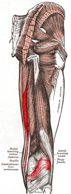 Muscles of the gluteal and posterior femoral regions (semimembranosus labeled at bottom left) | |
| Details | |
| Origin | Ischial tuberosity |
| Insertion | Medial condyle of tibia |
| Artery | Profunda femoris and gluteal arteries |
| Nerve | Tibial part of sciatic nerve (L5, S1 and S2) |
| Actions | Extension of hip and flexion of knee |
| Antagonist | Quadriceps muscle and Tensor fasciae latae |
| Identifiers | |
| Latin | Musculus semimembranosus |
| TA | A04.7.02.036 |
| FMA | 22438 |
| Anatomical terms of muscle | |
Structure
The semimembranosus, so called from its membranous tendon of origin, is situated at the back and medial side of the thigh.
Its origin is the superolateral aspect of the ischial tuberosity and it inserts on the medial condyle and nearby margin of tibia; intercondylar fossa of femur and medial condyle of femur; and the ligament of the popliteal region (at the back of the knee). It arises by a thick tendon from the upper and outer impression on the ischial tuberosity, above and medial to the biceps femoris and semitendinosus.
The tendon of origin expands into an aponeurosis, which covers the upper part of the anterior surface of the muscle; from this aponeurosis, muscular fibers arise, and converge to another aponeurosis which covers the lower part of the posterior surface of the muscle and contracts into the tendon of insertion.
It is inserted mainly into the horizontal groove on the posterior medial aspect of the medial condyle of the tibia.
The semimembranosus is wider, flatter, and deeper than the semitendinosus (with which it shares very close insertion and attachment points).
The tendon of insertion gives off certain fibrous expansions: one, of considerable size, passes upward and laterally to be inserted into the posterior lateral condyle of the femur, forming part of the oblique popliteal ligament of the knee-joint; a second is continued downward to the fascia which covers the popliteus muscle; while a few fibers join the medial collateral ligament of the joint and the fascia of the leg.
The muscle overlaps the upper part of the popliteal vessels.
Innervation
The semimembranosus is innervated by the tibial part of the sciatic nerve. The sciatic nerve consists of the anterior divisions of ventral nerve roots from L4 through S3. These nerve roots are part of the larger nerve network–the sacral plexus.[1] The tibial part of the sciatic nerve is also responsible for innervation of semitendinosus and the long head of biceps femoris.
Variation
It may be reduced or absent, or double, arising mainly from the sacrotuberous ligament and giving a slip to the femur or adductor magnus.
Function
The semimembranosus helps to extend (straighten) the hip joint and flex (bend) the knee joint.
It also helps to medially rotate the knee: the tibia medially rotates on the femur when the knee is flexed. It medially rotates the femur when the hip is extended. The muscle can also aid in counteracting the forward bending at the hip joint.[1]
Additional images
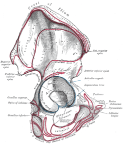 Right hip bone. External surface.
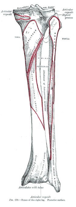 Bones of the right leg. Posterior surface.
 The popliteal, posterior tibial, and peroneal arteries.
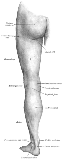 Back of left lower extremity.
 Semimembranosus muscle
 Semimembranosus muscle
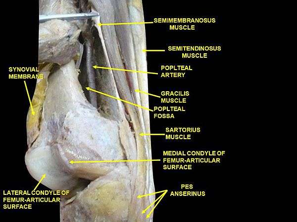 Muscles of thigh. Lateral view.
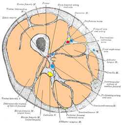
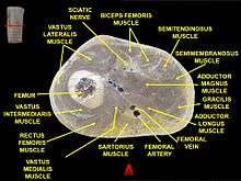 Muscles of thigh. Cross section.
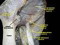 Muscles of thigh. Anterior views.
See also
References
This article incorporates text in the public domain from page 479 of the 20th edition of Gray's Anatomy (1918)
- Saladin, Kenneth S. Anatomy & Physiology: the unity of form and function. 5th ed. New York: McGraw-Hill, 2010. Print.
External links
| Wikimedia Commons has media related to Semimembranosus muscle. |
- Anatomy photo:14:st-0408 at the SUNY Downstate Medical Center
- Anatomy figure: 14:01-07 at Human Anatomy Online, SUNY Downstate Medical Center - "Muscles (hamstrings) of the posterior compartment of the thigh."
- Anatomy figure: 14:02-06 at Human Anatomy Online, SUNY Downstate Medical Center - "Muscles that form the superficial boundaries of the popliteal fossa."
- knee/surface/surface4 at the Dartmouth Medical School's Department of Anatomy
- PTCentral
| Authority control |
|---|