Subfornical organ
The subfornical organ (SFO) is one of the circumventricular organs of the brain.[1][2] Its name comes from its location on the ventral surface of the fornix near the interventricular foramina (foramina of Monro), which interconnect the lateral ventricles and the third ventricle. Like all circumventricular organs, the subfornical organ is well-vascularized, and like all circumventricular organs except the subcommissural organ, some SFO capillaries have fenestrae, which increase capillary permeability.[1][3][4] The SFO is considered a sensory circumventricular organ because it is responsive to a wide variety of hormones and neurotransmitters, as opposed to secretory circumventricular organs, which are specialized in the release of certain substances.[1][4]
| Subfornical organ | |
|---|---|
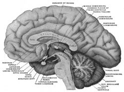 Medial aspect of a brain sectioned in the median sagittal plane. (Subfornical organ not labeled, but fornix and foramen of Monro are both labeled near the center.) | |
| Details | |
| Identifiers | |
| Latin | organum subfornicale |
| MeSH | D013356 |
| NeuroLex ID | nlx_anat_100314 |
| TA | A14.1.08.412 A14.1.09.449 |
| FMA | 75260 |
| Anatomical terms of neuroanatomy | |
Anatomy
As noted above, capillaries in some subregions within the SFO are fenestrated,[5] and thus lack a blood-brain barrier. All circumventricular organs except the subcommissural organ contain fenestrated capillaries,[2] a feature that distinguishes them from most other parts of the brain.[6] The SFO can be divided into six anatomical zones based on its capillary topography: two zones in the coronal plane and four zones in the sagittal plane.[3] The central zone is composed of the glial cells, neuronal cell bodies and a high density of fenestrated capillaries.[7] Conversely, the rostral and caudal areas have a lower density of capillaries[7] and are mostly made of nerve fibers, with fewer neurons and glial cells seen in this area. Functionally, however, the SFO may be viewed in two portions, the dorsolateral peripheral division, and the ventromedial core segment.[8]
The subfornical organ contains endothelin receptors mediating vasoconstriction and high rates of glucose metabolism mediated by calcium channels.[9]
General function
The subfornical organ is active in many bodily processes,[1] including, but not limited to, osmoregulation,[8] cardiovascular regulation,[8] and energy homeostasis.[10] Most of these processes involve fluid balance through the control of the release of certain hormones, particularly angiotensin or vasopressin.
Cardiovascular regulation
The subfornical organ's impact on the cardiovascular system is mostly mediated through its influence on fluid balance.[1] The SFO plays a role in vasopressin regulation. Vasopressin is a hormone that, when bound to receptors in the kidneys, increases water retention in the cardiovascular system by decreasing the amount of fluid transferred from the blood to the urine by the kidneys. This regulation of blood volume affects other aspects of the cardiovascular system. Increased or decreased blood volume influences blood pressure, which is regulated by baroreceptors, and can in turn affect the strength of ventricular contraction in the heart, although heart rate is generally not affected by blood volume. Additional research has demonstrated that the subfornical organ may be an important intermediary through which leptin acts to maintain blood pressure within normal physiological limits via descending autonomic pathways associated with cardiovascular control.[1]
SFO neurons have also been experimentally shown to send efferent projections to regions involved in cardiovascular regulation including the lateral hypothalamus, with fibers terminating in the supraoptic (SON) and paraventricular (PVN) nuclei, and the anteroventral 3rd ventricle (AV3V) with fibers terminating in the OVLT and the median preoptic area.[11]
Appetite and energy homeostasis
The subfornical organ has also been shown to have a significant impact on appetite. These mechanisms are not as clear as the neural mechanisms by which the SFO regulates fluid balance; however the most prevalent theory links the SFO's role in appetite control to its influence on energy, particularly glucose consumption. Recent study has focused on the SFO as an area particularly important in the regulation of energy. The observation that subfornical neurons are perceptive of a wide range of circulating energy balance signals and that electrical stimulation of the SFO in rats resulted in food intake supports the SFO’s importance in energy homeostasis.[10] Additionally, it is assumed that the SFO is the lone forebrain structure capable of constant monitoring of circulating concentrations of glucose, due to its lack of a blood-brain barrier.[10] This responsiveness to glucose again serves to solidify the SFO’s integral role as a regulator of energy homeostasis.[10]
Relationship with other circumventricular organs
Other circumventricular organs are the area postrema in the brainstem and the OVLT.[1][6]
The OVLT and SFO are both interconnected with the nucleus medianus, and together these three structures comprise the so-called "AV3V" region - the region anterior and ventral to the third ventricle.[1] The AV3V region is important in the regulation of fluid and electrolyte balance, by controlling thirst, sodium excretion, blood volume regulation, and vasopressin secretion.[11]
The SFO is outside the blood–brain barrier, and so neuronal hormone receptors in this region can respond to factors present in the systemic circulation.[1][7] The circumventricular organs express high density of Glucagon-like peptide 1 (GLP-1)receptors and participate in the central regulation of energy intake.
Hormones and receptors
Neurons in the subfornical organ have receptors for many hormones that circulate in the blood but which do not cross the blood–brain barrier,[1] including angiotensin, atrial natriuretic peptide, endothelin and relaxin. The role of the SFO in angiotensin regulation is particularly important, as it is involved in communication with the nucleus medianus (also called the median preoptic nucleus). Some neurons in the SFO are osmoreceptors, being sensitive to the osmotic pressure of the blood. These neurons project to the supraoptic nucleus and paraventricular nucleus to regulate the activity of vasopressin-secreting neurons. These neurons also project to the nucleus medianus which is involved in controlling thirst. Thus, the subfornical organ is involved in fluid balance.
Other important hormones have been shown to excite the SFO, specifically serotonin, carbamylcholine (carbachol), and atropine. These neurotransmitters however seem to have an effect on deeper areas of the SFO than angiotensin, and antagonists of these hormones have been shown to also primarily effect the non-superficial regions of the SFO (other than atropine antagonists, which showed little effects). In this context, the superficial region is considered to be 15-55μm deep into the SFO, and the "deep" region anything below that.
From these reactions to certain hormones and other molecules, a model of the neuronal organization of the SFO is suggested in which angiotensin-sensitive neurons lying superficially are excited by substances borne by blood or cerebrospinal fluid, and synapse with deeper carbachol-sensitive neurons. The axons of these deep neurons pass out of the SFO in the columns and body of the fornix. Afferent fibers from the body and columns of the fornix polysynaptically excite both superficial and deep neurons. A recurrent inhibitory circuit is suggested on the output path.[11]
Genetics
The expression of various genes in the subfornical organ have been studied. For example, it was seen that water deprivation in rats led to an upregulation of the mRNA that codes for angiotensin II receptors, allowing for a lower angiotensin concentration in the blood that produce the "thirst" response. It also has been observed to be a site of thyroid transcription factor 1 (TTF1) production, a protein generally produced in the hypothalamus.[12]
Pathology
Hypertension
Hypertension, or high blood pressure, is highly affected by the concentration of angiotensin. Injection of angiontensin has actually been long used to induce hypertension in animal test models to study the effects of various therapies and medications. In such experiments, it has been observed that an intact and functioning subfornical organ limits the increase in mean arterial pressure due to the increased angiotensin.[13]
Dehydration
As stated above, angiotensin receptors (AT1) have been shown to be upregulated due to water deprivation. These AT1 receptors have also shown an increased bonding with circulating angiotensin after water deprivation. These findings could indicate some sort of morphological change in the AT1 receptor, likely due to some signal protein modification of the AT1 receptor at a non-bonding site, leading to an increased affinity of the AT1 receptor for angiotensin bonding.[14]
Research
Feeding
Although generally viewed primarily as having roles in homeostasis and cardiovascular regulation, the subfornical organ has been thought to control feeding patterns through taking inputs from the bloodstream (various peptides indicating satiety) and then stimulating hunger. It has been shown to induce drinking in rats as well as eating.
One study looks at different stimulation current values, to determine if this has an effect on the amount of feeding that occurs. The rats studied were separated into three groups: rats with electrodes in their subfornical organ with no current passing through (sham), rats with stimulated subfornical organs, and rats with areas other than the subfornical organ stimulated. The group with stimulated subfornical organs was separated into groups with 100mA and 200mA stimulations. All rats were satiated (food and drink) before observations/stimulations were done, and were also monitored for general activity. The group with subfornical stimulation at 100mA drank an increased amount, but did not consume any additional food, and the group with 200mA consumed both more water and more food. All groups without subfornical organ stimulation did not eat or drink at all.[15]
Other studies look specifically at drinking, as the SFO is known to have an important role in fluid balance. One such study looked into the connection between the SFO and the median preoptic nucleus. Rats with both partially or fully severed connections to the median preoptic nucleus showed a significantly decreased tendency to drink water when compared to the control group. Then when angiotensin was injected subcutaneously, drinking incidence went back to original levels. These findings are consistent with a model that postulates that osmoreceptors and angiotensin receptors in the SFO send excitatory neural information to the median preoptic nucleus for the mobilization of thirst.[11]
References
- Gross, P. M; Weindl, A (1987). "Peering through the windows of the brain (review)". Journal of Cerebral Blood Flow & Metabolism. 7 (6): 663–72. doi:10.1038/jcbfm.1987.120. PMID 2891718.
- Oldfield BJ and Mckinley MJ (1995). Paxinos G (ed.). The Rat Nervous System. San Diego: Academic Press. pp. 391–403. ISBN 978-0-12-547635-5.CS1 maint: uses authors parameter (link)
- Sposito NM, Gross PM (1987). "Topography and morphometry of capillaries in the rat subfornical organ". J Comp Neurol. 260 (1): 36–46. doi:10.1002/cne.902600104. PMID 3597833.
- Miyata, S (2015). "New aspects in fenestrated capillary and tissue dynamics in the sensory circumventricular organs of adult brains". Frontiers in Neuroscience. 9: 390. doi:10.3389/fnins.2015.00390. PMC 4621430. PMID 26578857.
- Shaver SW, Sposito NM, Gross PM (1990). "Quantitative fine structure of capillaries in subregions of the rat subfornical organ". J Comp Neurol. 294 (1): 145–52. doi:10.1002/cne.902940111. PMID 2324330.
- Gross PM (1992). Circumventricular organ capillaries (review). Prog Brain Res. Progress in Brain Research. 91. pp. 219–33. doi:10.1016/S0079-6123(08)62338-9. ISBN 9780444814197. PMID 1410407.
- Gross PM (1991). "Morphology and physiology of capillary systems in subregions of the subfornical organ and area postrema". Can J Physiol Pharmacol. 69 (7): 1010–25. doi:10.1139/y91-152. PMID 1954559.
- Kawano, H.; Masuko, S. (2010). "Region-specific projections from the subfornical organ to the paraventricular hypothalamic nucleus in the rat". Neuroscience. 169 (3): 1227–34. doi:10.1016/j.neuroscience.2010.05.065. PMID 20678996.
- Gross PM, Wainman DS, Chew BH, Espinosa FJ, Weaver DF (1993). "Calcium-mediated metabolic stimulation of neuroendocrine structures by intraventricular endothelin-1 in conscious rats". Brain Res. 606 (1): 135–42. doi:10.1016/0006-8993(93)91581-c. PMID 8461995.
- Medeiros, N., Dai, L., and Ferguson, A.V. (2012). Glucose-responsive neurons in the subfornical organ of the rat—a novel site for direct CNS monitoring of circulating glucose. Neuroscience 201, 157-165.
- Lind R. Wallace (1982). "Subfornical organ-median preoptic connections and drinking and pressor responses to angiotensin II". The Journal of Neuroscience. 2 (3): 1043–51. doi:10.1523/JNEUROSCI.02-08-01043.1982.
- Son YJ, Hur MK, Ryu BJ, Park SK (2003). "TTF-1, a homeodomain-containing transcription factor, participates in the control of body fluid homeostasis by regulating angiotensinogen gene transcription in the rat subfornical organ". The Journal of Biological Chemistry. 278 (29): 27043–52. doi:10.1074/jbc.M303157200. PMID 12730191.
- Bruner CA, Mangiopane ML, Fink GD (1985). "Subfornical organ. Does it protect against angiotensin II-induced hypertension in the rat?". Circ. Res. 56 (3): 462–6. doi:10.1161/01.res.56.3.462. PMID 3971518.
- Sanvitto G L, Johren 0, Hauser W, Saavedra J M (1997). "Water deprivation upregulates ANG II AT1 binding and mRNA in rat subfornical organ and anterior pituitary". Journal of Physiology.CS1 maint: multiple names: authors list (link)
- Smith P (2010). "Acute electrical stimulation of the subfornical organ induces feeding in satiated rats". Physiology and Behavior. 99 (4): 534–37. doi:10.1016/j.physbeh.2010.01.013. PMID 20096716.
External links
| Authority control |
|---|