Interthalamic adhesion
The interthalamic adhesion (also known as the intermediate mass or middle commissure) is a flattened band of tissue that connects both parts of the thalamus at their medial surfaces. The medial surfaces form the upper part of the lateral wall to the third ventricle.
| Interthalamic adhesion | |
|---|---|
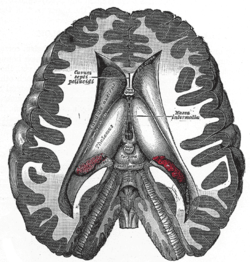 Dissection showing the ventricles of the brain. (Interthalamic adhesion labeled as Massa Intermedia at center right.) | |
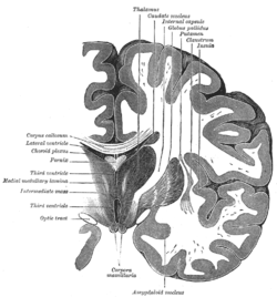 Coronal section of brain through intermediate mass of third ventricle. | |
| Details | |
| Part of | thalamus |
| Identifiers | |
| Latin | Adhaesio interthalamica |
| NeuroNames | 301 |
| NeuroLex ID | nlx_144100 |
| TA | A14.1.08.103 |
| FMA | 74869 |
| Anatomical terms of neuroanatomy | |
In mammals other than humans, it is a large structure. In humans it is only about one centimeter long, though in females it is larger by about 50%.[1] Sometimes it is in two parts and 20% to 30% of the time it is absent.
In 1889, a Portuguese anatomist by the name of Macedo examined 215 brains, showing that male humans are approximately twice as likely to lack an interthalamic adhesion as are female humans. He anecdotally attributed the finding to a "prevailing feature of people deprived of [the interthalamic adhesion] is to present in their psychical acts a remarkable precipitation, joined to a certain dysharmony between internal and external feelings".[2] Its absence is seen to be inconsequential.
The interthalamic adhesion contains nerve cells and nerve fibers; a few of the latter may cross the middle line, but most of them pass toward the middle line and then curve laterally on the same side. It is still uncertain whether the interthalamic adhesion contains fibers that cross the mid-line and for this reason it is inappropriate to call it a commissure.
The interthalamic adhesion is notably enlarged in patients with the type II Arnold-Chiari malformation.[3]
Additional images
 Thalamus
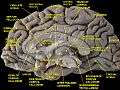 Medial surface of cerebral hemisphere.Medial view.Deep dissection. WRONG INFORMATION - NEED REVIEW
References
This article incorporates text in the public domain from the 20th edition of Gray's Anatomy (1918)
- Wiley Interscience
- REGIS OLRY AND DUANE E. HAINES, "Interthalamic Adhesion: Scruples About Calling a Spade a Spade?" Journal of the History of the Neurosciences, 14:116-118, 2005
- Wolpert, S. M.; Anderson, M; Scott, R. M.; Kwan, E. S.; Runge, V. M. (1987). "Chiari II malformation: MR imaging evaluation". American Journal of Roentgenology. 149 (5): 1033–42. doi:10.2214/ajr.149.5.1033. PMID 3499774.
External links
- Atlas image: n1a8p6 at the University of Michigan Health System
- Image at Harvard University
- Diagram at csuchico.edu (labeled as Massa intermedia)
- "Anatomy diagram: 13048.000-3". Roche Lexicon - illustrated navigator. Elsevier. Archived from the original on 2012-07-22.
| Authority control |
|---|