Hippocampus proper
The hippocampus proper refers to the actual structure of the hippocampus which is made up of four regions or subfields. The subfields CA1, CA2, CA3, and CA4 use the initials of cornu Ammonis, an earlier name of the hippocampus.
| Hippocampus proper | |
|---|---|
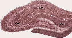 Diagram of hippocampal regions in a rat brain. | |
.png) Basic circuit of the hippocampus, shown using a modified drawing by Ramon y Cajal. DG: dentate gyrus. Sub: subiculum. EC: entorhinal cortex | |
| Details | |
| Identifiers | |
| Latin | hippocampus proprius, cornu Ammonis |
| NeuroNames | 182 |
| TA | A14.1.09.327 |
| FMA | 62493 |
| Anatomical terms of neuroanatomy | |
Structure
There are four regions in the hippocampus proper which form a neural circuit called the trisynaptic circuit.
CA1
CA1 is the first region in the hippocampal circuit, from which a major output pathway goes to layer V of the entorhinal cortex. Another significant output is to the subiculum.
CA2
CA2 is a small region located between CA1 and CA3. It receives some input from layer II of the entorhinal cortex via the perforant path. Its pyramidal cells are more like those in CA3 than those in CA1. It is often ignored due to its small size.
CA3
CA3 receives input from the mossy fibers of the granule cells in the dentate gyrus, and also from cells in the entorhinal cortex via the perforant path. The mossy fiber pathway ends in the stratum lucidum. The perforant path passes through the stratum lacunosum and ends in the stratum moleculare. There are also inputs from the medial septum and from the diagonal band of Broca which terminate in the stratum radiatum, along with commisural connections from the other side of the hippocampus.
The pyramidal cells in CA3 send some axons back to the dentate gyrus hilus, but they mostly project to regions CA2 and CA1 via the Schaffer collaterals. There are also a significant number of recurrent connections that terminate in CA3. Both the recurrent connections and the Schaffer collaterals terminate preferentially in the septal area in a dorsal direction from the originating cells. CA3 also sends a small set of output fibers to the lateral septum.
The region is conventionally divided into three divisions. CA3a is the part of the cell band that is most distant from the dentate (and closest to CA1). CA3b is the middle part of the band nearest to the fimbria and fornix connection. CA3c is nearest to the dentate, inserting into the hilus. CA3 overall, has been considered to be the “pacemaker” of the hippocampus. Much of the synchronous bursting activity associated with interictal epileptiform activity appears to be generated in CA3. Its excitatory collateral connectivity seems to be mostly responsible for this. CA3 uniquely, has pyramidal cell axon collaterals that ramify extensively with local regions and make excitatory contacts with them. CA3 has been implicated in a number of working theories on memory and hippocampal learning processes. Slow oscillatory rhythms (theta-band; 3–8 Hz) are cholinergically driven patterns that depend on coupling of interneurons and pyramidal cell axons via gap junctions, as well as glutaminergic (excitatory) and GABAergic (inhibitory) synapses. Sharp EEG waves seen here are also implicated in memory consolidation.[1]
CA4
CA4 is often called the hilus or hilar region if considered as a part of the dentate gyrus. Unlike the pyramidal neurons in CA1 and CA3, the neurons here include mossy cells that primarily receive inputs from the granule cells in the dentate gyrus in the form of mossy fibers.[2][3] They also receive a small number of connections from pyramidal cells in CA3. They, in turn, project back into the dentate gyrus at distant septotemporal levels.
Additional images
.jpg) Hippocampus
Hippocampus
References
- Jerome Engel TAP, ed. Epilepsy: A Comprehensive Textbook in Three Volumes. Philadelphia, PA: Lippincott Williams & Wilkins; 2008
- Lorente de Nó, R (1934). "Studies on the structure of the cerebral cortex. Continuation of the study of the ammonic system". J. Psychol. Neurol. 46: 113–177.
- Amaral, DG (1978). "A Golgi study of cell types in the hilar region of the hippocampus in the rat". J. Comp. Neurol. 182 (5): 851–914. doi:10.1002/cne.901820508. PMID 730852.
| Authority control |
|---|