Medial lemniscus
The medial lemniscus, also known as Reil's band or Reil's ribbon, is a large ascending bundle of heavily myelinated axons that decussate in the brainstem, specifically in the medulla oblongata. The medial lemniscus is formed by the crossings of the internal arcuate fibers. The internal arcuate fibers are composed of axons of nucleus gracilis and nucleus cuneatus. The axons of the nucleus gracilis and nucleus cuneatus in the medial lemniscus have cell bodies that lie contralaterally.
| Medial lemniscus | |
|---|---|
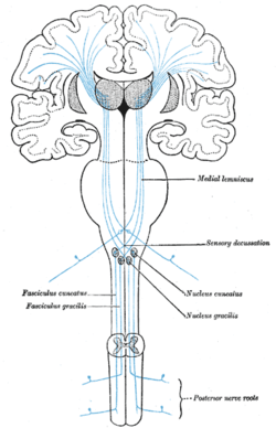 The sensory tract. (Medial lemniscus labeled at top right.) | |
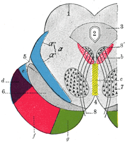 Coronal section through mid-brain. ("e" is Portion of medial lemniscus, which runs to the lentiform nucleus and insula. "a’" is also the medial lemniscus.) | |
| Details | |
| Identifiers | |
| Latin | lemniscus medialis |
| NeuroLex ID | birnlex_887 |
| TA | A14.1.04.111 A14.1.08.672 A14.1.06.207 |
| FMA | 83675 |
| Anatomical terms of neuroanatomy | |
The medial lemniscus is part of the dorsal column–medial lemniscus pathway, which ascends from the skin to the thalamus,[1] which is important for somatosensation from the skin and joints, therefore, lesion of the medial lemnisci causes an impairment of vibratory and touch-pressure sense.
Etymology
Lemniscus means "ribbon", so named because the medial lemniscus "spirals" or "turns" as it ascends.
Path
After neurons carrying proprioceptive or fine touch information synapse at the gracile and cuneate nuclei, axons from secondary neurons decussate at the level of the medulla and travel up the brainstem as the medial lemniscus on the contralateral (opposite) side. It is part of the posterior column-medial lemniscus pathway, which transmits touch, vibration sense, as well as the pathway for proprioception.
The medial lemniscus carries axons from most of the body and synapses in the ventral posterolateral nucleus of the thalamus, at the level of the mamillary bodies. Sensory axons transmitting information from the head and neck via the trigeminal nerve synapse at the ventral posteromedial nucleus of the thalamus.
Location through the brainstem
- The cuneate and gracile nuclei reside at the closed (lower) medulla, so the lemniscus is not formed at this level. Fibres from these nuclei will pass to the contralateral side of the brainstem, as the internal arcuate fibres.
- At the open medulla (further up the brainstem), the medial lemniscus contains axons from the trigeminal nerve (which supplies the head region), as well as the arms and legs. It sits very close to the midline, at the same orientation of the midline, with head fibres more dorsal (closer to the back), towards the fourth ventricle.
- By mid-pons, the medial lemniscus has rotated. Fibres from the head are medial, fibres from the leg are lateral.
- The orientation in the midbrain is similar to that in the pons.
Additional images
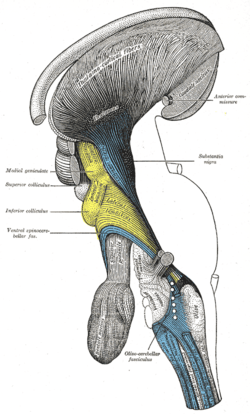 Deep dissection of brain-stem. Lateral view.
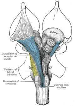 Deep dissection of brain-stem. Ventral view.
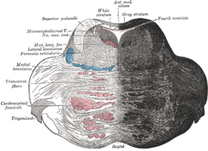 Coronal section of the pons, at its upper part.
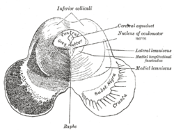 Transverse section of mid-brain at level of inferior colliculi.
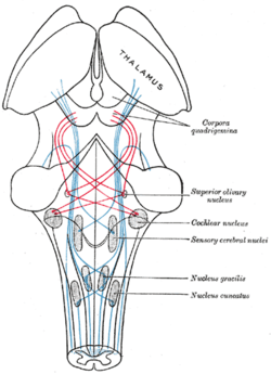 Scheme showing the course of the fibers of the lemniscus; medial lemniscus in blue, lateral in red.
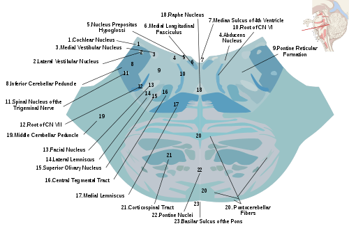 Horizontal section through the lower part of the pons. The medial lemniscus is labeled #17.
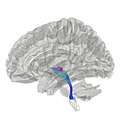 Tractography showing medial lemniscus
References
- Kamali A, Kramer LA, Butler IJ, Hasan KM. Diffusion tensor tractography of the somatosensory system in the human brainstem: initial findings using high isotropic spatial resolution at 3.0 T. Eur Radiol. 2009 19:1480-8. doi:10.1007/s00330-009-1305-x.
External links
| Authority control |
|---|