Median eminence
The median eminence, part of the inferior boundary of the hypothalamus in the brain, is attached to the infundibulum. The median eminence is a small swelling on the tuber cinereum, posterior to and atop the pituitary stalk; it lies in the area roughly bounded on its posterolateral region by the cerebral peduncles, and on its anterolateral region by the optic chiasm.
| Median eminence | |
|---|---|
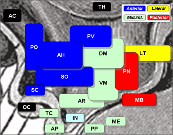 Median eminence is 'ME', at bottom-center, in light-green | |
| Details | |
| Identifiers | |
| Latin | eminentia mediana hypothalami |
| MeSH | D008473 |
| NeuroNames | 402 |
| NeuroLex ID | birnlex_925 |
| TA | A14.1.08.409 |
| FMA | 74634 |
| Anatomical terms of neuroanatomy | |
As one of the seven areas of the brain devoid of a blood–brain barrier,[1] the median eminence is a circumventricular organ having permeable capillaries.[2][3][4][5] Its main function is as a gateway for release of hypothalamic hormones,[6] although it does share contiguous perivascular spaces with the adjacent hypothalamic arcuate nucleus, indicating a potential sensory role.[3][7]
Physiology
The median eminence is a part of the hypothalamus from which regulatory hormones are released.[1][6] It is integral to the hypophyseal portal system, which connects the hypothalamus with the pituitary gland. The pars nervosa (part of the posterior pituitary gland) is continuous with the median eminence of the hypothalamus via the infundibular stalk. Parvocellular neurons from the hypothalamus terminate in the median eminence of the hypothalamus.
The median eminence is the structure where secretions of the hypothalamus (releasing and inhibiting regulatory hormones, known as "hypophysiotropic hormones") collect before entering the portal system emptying into the general circulation.[1][6] Such hypophysiotropic hormones include: CRF (corticotropin-releasing factor), GnRH (gonadotropin-releasing hormone), TRH (thyrotropin-releasing hormone), GHRH (growth hormone-releasing hormone), and DA (dopamine).[6] These hypophysiotropic hormones stimulate or inhibit the release of hormones from the anterior pituitary.[6] Further, anatomical evidence exists for bidirectional communication between the median eminence and the arcuate and ventromedial nucleus of the hypothalamus.[3][7]
References
- Gross PM, Weindl A (December 1987). "Peering through the windows of the brain". Journal of Cerebral Blood Flow and Metabolism. 7 (6): 663–72. doi:10.1038/jcbfm.1987.120. PMID 2891718.
- Scott DE, Pepe GJ (July 1987). "The fetal baboon median eminence as a circumventricular organ: I. Transmission electron microscopy". Brain Research Bulletin. 19 (1): 87–94. doi:10.1016/0361-9230(87)90170-5. PMID 3651843.
- Gross PM (1992). "Circumventricular organ capillaries". Progress in Brain Research. 91: 219–33. doi:10.1016/S0079-6123(08)62338-9. PMID 1410407.
- Johnson AK, Gross PM (May 1993). "Sensory circumventricular organs and brain homeostatic pathways". FASEB Journal. 7 (8): 678–86. doi:10.1096/fasebj.7.8.8500693. PMID 8500693.
- Ganong WF (2000). "Circumventricular organs: definition and role in the regulation of endocrine and autonomic function". Clinical and Experimental Pharmacology & Physiology. 27 (5–6): 422–7. doi:10.1046/j.1440-1681.2000.03259.x. PMID 10831247.
- Palkovits M (1984). "Neuropeptides in the hypothalamo-hypophyseal system: lateral retrochiasmatic area as a common gate for neuronal fibers towards the median eminence". Peptides. 5 Suppl 1: 35–9. doi:10.1016/0196-9781(84)90262-6. PMID 6148739.
- Shaver SW, Pang JJ, Wainman DS, Wall KM, Gross PM (March 1992). "Morphology and function of capillary networks in subregions of the rat tuber cinereum". Cell and Tissue Research. 267 (3): 437–48. doi:10.1007/BF00319366. PMID 1571958.
| Authority control |
|---|