Phthisis bulbi
Phthisis bulbi is a shrunken,[1] non-functional eye. It may result from severe eye disease, inflammation[2] or injury, or it may represent a complication of eye surgery.[3] Treatment options include insertion of a prosthesis, which may be preceded by enucleation of the eye.[4][5]
| Phthisis bulbi | |
|---|---|
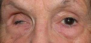 | |
| Phthisis bulbi of the right eye due to complication of eye surgery | |
| Specialty | Ophthalmology |
| Symptoms | Shrunken eye with little or no function |
| Causes | Eye surgery |
| Risk factors | Eye injury, Eye surgery, eye disease |
| Prevention | By treating the condition before the eye goes to pthisis |
| Treatment | Surgery |
| Prognosis | Usually permanent blindness in affected eye |
| Deaths | 0 |
Symptoms
The affected eye is shrunken, and has little to no vision. The intraocular pressure in the affected eye is very low or nonexistent. The layers in the eye may be fused together, thickened, or edematous. The eyelids may be glued shut. The eye may be soft when palpated.[6] Under a microscope there may be deposits of calcium or bone, and the cornea is often affected by cataracts.[7]
Causes
It can be caused by injury, including burns to the eye, or long-term eye disease or inflammation. End-stage glaucoma can cause it. It can often complicate eye surgery.[6] Other common causes include cancer, retinal detachment, vascular lesions, infection, and inflammation.[7]
Treatment
Treatment for the affected eye is often futile. Usually, treatment is to end the pain in the affected eye and for cosmetic purposes, not to restore vision.[7] It can be removed, a procedure called enucleation of the eye. Sometimes, though, it is possible to transplant only parts of the eye, and some vision can be restored.[6]
References
- Dornblüth, von Willibald Pschyrembel. Gegr. von Otto (1977). Klinisches Wörterbuch : mit klinischen Syndromen und einem Anhang Nomina Anatomica (253., um einen Anh. 'Nomina anatomica' erw. Aufl. ed.). Berlin [u.a.]: de Gruyter. ISBN 978-3-11-007018-7.
- Hui, JI (September 2010). "Outcomes of orbital implants after evisceration and enucleation in patients with endophthalmitis". Current Opinion in Ophthalmology. 21 (5): 375–9. doi:10.1097/ICU.0b013e32833b7a56. PMID 20489621.
- Apple, DJ; Jones, GR; Reidy, JJ; Loftfield, K (Sep–Oct 1985). "Ocular perforation and phthisis bulbi secondary to strabismus surgery". Journal of Pediatric Ophthalmology and Strabismus. 22 (5): 184–7. PMID 4045647.
- Cote, RE; Haddad, SE (1990). "Fitting a prosthesis over phthisis bulbi or discolored blind eyes". Advances in Ophthalmic Plastic and Reconstructive Surgery. 8: 136–45. PMID 2248703.
- Soares, IP; França, VP (May 2010). "Evisceration and enucleation". Seminars in Ophthalmology. 25 (3): 94–7. doi:10.3109/08820538.2010.488575. PMID 20590419.
- D'Amico, Donald J.; Dohlman, Claes H. (1999-01-01). "Can an Eye in Phthisis Be Rehabilitated? A Case of Improved Vision With 1-Year Follow-up". Archives of Ophthalmology. 117 (1): 123–124. doi:10.1001/archopht.117.1.123. ISSN 0003-9950.
- Tripathy, Koushik; Chawla, Rohan; Temkar, Shreyas; Sagar, Pradeep; Kashyap, Seema; Pushker, Neelam; Sharma, Yog Raj (2018). "Phthisis Bulbi-a Clinicopathological Perspective". Seminars in Ophthalmology. 33 (6): 788–803. doi:10.1080/08820538.2018.1477966. ISSN 1744-5205. PMID 29902388.