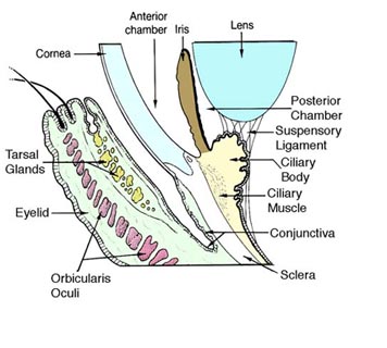
Conjunctiva
The conjunctiva is a tissue that lines the inside of the eyelids and covers the sclera (the white of the eye). It is composed of unkeratinized, stratified squamous epithelium with goblet cells, and stratified columnar epithelium. The conjunctiva is highly vascularised, with many microvessels easily accessible for imaging studies.
| Conjunctiva | |
|---|---|
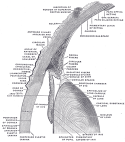 The upper half of a sagittal section through the front of the eyeball. (Label for 'Conjunctiva' visible at center-left) | |
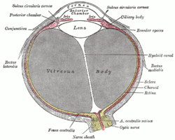 Horizontal section of the eyeball. (Conjunctiva labeled at upper left) | |
| Details | |
| Part of | Eye |
| Artery | lacrimal artery, anterior ciliary arteries |
| Nerve | supratrochlear nerve |
| Identifiers | |
| Latin | tunica conjunctiva |
| MeSH | D003228 |
| TA | A15.2.07.047 |
| FMA | 59011 |
| Anatomical terminology | |
Structure
The conjunctiva is typically divided into three parts:
| Part | Area |
|---|---|
| Palpebral or tarsal conjunctiva | Lines the eyelids |
| Bulbar or ocular conjunctiva | Covers the eyeball, over the anterior sclera: This region of the conjunctiva is tightly bound to the underlying sclera by Tenon's capsule and moves with the eyeball movements. The average thickness of the bulbar conjuntival membrane is 33 microns.[1] |
| Fornix conjunctiva | Forms the junction between the bulbar and palpebral conjunctivas: It is loose and flexible, allowing the free movement of the lids and eyeball.[2] |
Blood supply
Blood to the bulbar conjunctiva is primarily derived from the ophthalmic artery. The blood supply to the palpebral conjunctiva (the eyelid) is derived from the external carotid artery. However, the circulations of the bulbar conjunctiva and palpebral conjunctiva are linked, so both bulbar conjunctival and palpebral conjunctival vessels are supplied by both the ophthalmic artery and the external carotid artery, to varying extents.[3]
Nerve supply
Sensory innervation of the conjunctiva is divided into four parts:[4]
| Area | Nerve |
|---|---|
| Superior | |
| Inferior | Infraorbital nerve |
| Lateral | Lacrimal nerve (with contribution from zygomaticofacial nerve) |
| Circumcorneal | Long ciliary nerves |
Microanatomy
The conjunctiva consists of unkeratinized, both stratified squamous and stratified columnar epithelium, with interspersed goblet cells.[5] The epithelial layer contains blood vessels, fibrous tissue, and lymphatic channels.[5] Accessory lacrimal glands in the conjunctiva constantly produce the aqueous portion of tears.[5] Additional cells present in the conjunctival epithelium include melanocytes, T and B cell lymphocytes.[5]
Function
The conjunctiva helps lubricate the eye by producing mucus and tears, although a smaller volume of tears than the lacrimal gland.[6] It also contributes to immune surveillance and helps to prevent the entrance of microbes into the eye.
Clinical significance
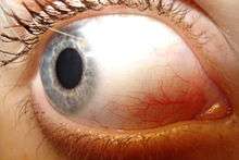
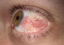
Disorders of the conjunctiva and cornea are common sources of eye complaints, in particular because the surface of the eye is exposed to various external influences and is especially susceptible to trauma, infections, chemical irritation, allergic reactions, and dryness.
- The conjunctival microvascular hemodynamics are affected by diabetic retinopathy (DR), hence can be useful for DR diagnosis and monitoring,[7] and discriminating stages of DR.[8]
- Type II diabetes is associated with conjunctival hypoxia,[9] increased average blood vessel diameter, and capillary loss.[10][11][12]
- Sickle-cell anemia is associated with blood vessel sludging, altered blood flow and blood vessel diameter, and capillary micro-haemorrhages.[13][14][15]
- Hypertension is associated with an increase in the tortuosity of bulbar conjunctival blood vessels and capillary and arteriole loss.[16][17]
- Carotid artery occlusion is associated with slower conjunctival blood flow and apparent capillary loss.[3]
- With age, the conjunctiva can stretch and loosen from the underlying sclera, leading to the formation of conjunctival folds, a condition known as conjunctivochalasis.[18][19]
- The conjunctiva can be affected by tumors which can be benign, pre-malignant or malignant.[20]
- Leptospirosis, an infection with Leptospira, can cause conjunctival suffusion, which is characterized by chemosis, and redness without exudates.
Bulbar conjunctival microvasculature
Vessel morphology
The bulbar conjunctival microvasculature contains arterioles, meta-arterioles, venules, capillaries, and communicating vessels. Vessel morphology varies greatly between subjects and even between regions of the individual eyes. In some subjects, arterioles and venules can be seen to run parallel with each other. Paired arterioles are generally smaller than corresponding venules.[21] The average bulbar conjunctival vessel has been reported to be 15.1 microns, which reflects the high number of small capillaries, which are typically <10 microns in diameter.[22]
Blood oxygen dynamics
The bulbar conjunctival microvasculature is in close proximity to ambient air, thus oxygen diffusion from ambient air strongly influences their blood oxygen saturation. Because of oxygen diffusion, hypoxic bulbar conjunctival vessels will rapidly reoxygenate (in under 10 seconds) when exposed to ambient air (i.e. when the eyelid is open). Closing the eyelid stops this oxygen diffusion by placing a barrier between the bulbar conjunctival microvessels and ambient air.[23]
Blood vessel imaging methods
The bulbar conjunctival microvessels are typically imaged with a high-magnification slit lamp with green filters.[24][25][26] With such high-magnification imaging systems, it is possible to see groups of individual red blood cells flowing in vivo.[24] Fundus cameras may also be used for low-magnification wide field-of-view imaging of the bulbar conjunctival microvasculature. Modified fundus cameras have been used to measure conjunctival blood flow [27] and to measure blood oxygen saturation.[23] Fluorescein angiography has been used to study the blood flow of the bulbar conjunctiva and to differentiate the bulbar conjunctival and episcleral microcirculation.[28][29][30]
Vasodilation
The bulbar conjunctival microvasculature is known to dilate in response to several stimuli and external conditions, including allergens (e.g. pollen),[31] temperature,[32] time-of-day,[32] contact-lens wear,[12] and acute mild hypoxia.[23] Bulbar conjunctival vasodilation has also been shown to correlate changes in emotional state.[33]
Type 2 diabetes is associated with an increase in average bulbar conjunctival vessel diameter and capillary loss.[10][11] Sickle-cell anemia is associated with altered average vessel diameter.[13]
See also
Additional images
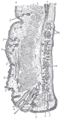 Sagittal section through the upper eyelid.
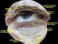 Extrinsic eye muscle. Nerves of orbita. Deep dissection.
References
- Efron, Nathan; Al-Dossari, Munira; Pritchard, Nicola (2009-05-01). "In vivo confocal microscopy of the bulbar conjunctiva". Clinical & Experimental Ophthalmology. 37 (4): 335–344. doi:10.1111/j.1442-9071.2009.02065.x. ISSN 1442-9071. PMID 19594558.
- Eye, human Encyclopædia Britannica
- PAVLOU AT; WOLFF HG (1959-07-01). "THe bulbar conjunctival vessels in occlusion of the internal carotid artery". Archives of Internal Medicine. 104 (1): 53–60. doi:10.1001/archinte.1959.00270070055007. ISSN 0888-2479.
- "Table 1: Summary of sensory nerve supply". Archived from the original on February 14, 2013. Retrieved July 31, 2016.
- Goldman, Lee. Goldman's Cecil Medicine (24th ed.). Philadelphia: Elsevier Saunders. p. 2426. ISBN 978-1437727883.
- London Place Eye Center (2003). Conjunctivitis Archived 2004-08-08 at the Wayback Machine. Retrieved July 25, 2004.
- Khansari, Maziyar M.; Wanek, Justin; Tan, Michael; Joslin, Charlotte E.; Kresovich, Jacob K.; Camardo, Nicole; Blair, Norman P.; Shahidi, Mahnaz (7 April 2017). "Assessment of Conjunctival Microvascular Hemodynamics in Stages of Diabetic Microvasculopathy". Scientific Reports. 7: 45916. Bibcode:2017NatSR...745916K. doi:10.1038/srep45916. PMC 5384077. PMID 28387229.
- Khansari, Maziyar M.; O’Neill, William; Penn, Richard; Chau, Felix; Blair, Norman P.; Shahidi, Mahnaz (1 July 2016). "Automated fine structure image analysis method for discrimination of diabetic retinopathy stage using conjunctival microvasculature images". Biomedical Optics Express. 7 (7): 2597–2606. doi:10.1364/BOE.7.002597. ISSN 2156-7085. PMC 4948616. PMID 27446692.
- Isenberg, S. J.; McRee, W. E.; Jedrzynski, M. S. (1986-10-01). "Conjunctival hypoxia in diabetes mellitus". Investigative Ophthalmology & Visual Science. 27 (10): 1512–1515. ISSN 0146-0404. PMID 3759367.
- Fenton, B. M.; Zweifach, B. W.; Worthen, D. M. (1979-09-01). "Quantitative morphometry of conjunctival microcirculation in diabetes mellitus". Microvascular Research. 18 (2): 153–166. doi:10.1016/0026-2862(79)90025-6. ISSN 0026-2862. PMID 491983.
- Ditzel, Jørn (1967-01-12). "The in Vivo Reactions of the Small Blood Vessels to Diabetes Mellitus". Acta Medica Scandinavica. 182 (S476): 123–134. doi:10.1111/j.0954-6820.1967.tb12691.x. ISSN 0954-6820.
- Cheung, A. T.; Ramanujam, S.; Greer, D. A.; Kumagai, L. F.; Aoki, T. T. (2001-10-01). "Microvascular abnormalities in the bulbar conjunctiva of patients with type 2 diabetes mellitus". Endocrine Practice. 7 (5): 358–363. doi:10.4158/EP.7.5.358. ISSN 1530-891X. PMID 11585371.
- Fink, A I (1968-01-01). "Vascular changes in the bulbar conjunctiva associated with sickle-cell disease: some observations on fine structure". Transactions of the American Ophthalmological Society. 66: 788–826. ISSN 0065-9533. PMC 1310317. PMID 5720854.
- Isenberg, S. J.; McRee, W. E.; Jedrzynski, M. S.; Gange, S. N.; Gange, S. L. (1987-01-01). "Effects of sickle cell anemia on conjunctival oxygen tension and temperature". Archives of Internal Medicine. 147 (1): 67–69. doi:10.1001/archinte.147.1.67. ISSN 0003-9926. PMID 3800533.
- Wanek, Justin; Gaynes, Bruce; Lim, Jennifer I.; Molokie, Robert; Shahidi, Mahnaz (2013-08-01). "Human bulbar conjunctival hemodynamics in hemoglobin SS and SC disease". American Journal of Hematology. 88 (8): 661–664. doi:10.1002/ajh.23475. ISSN 1096-8652. PMC 4040222. PMID 23657867.
- Harper, Robert N.; Moore, Michael A.; Marr, Melissa C.; Watts, L. Earl; Hutchins, Phillip M. (1978-11-01). "Arteriolar rarefaction in the conjunctiva of human essential hypertensives". Microvascular Research. 16 (3): 369–372. doi:10.1016/0026-2862(78)90070-5.
- Lee, R. E. (1955-08-01). "Anatomical and physiological aspects of the capillary bed in the bulbar conjunctiva of man in health and disease". Angiology. 6 (4): 369–382. doi:10.1177/000331975500600408. ISSN 0003-3197. PMID 13275744.
- "Conjunctivochalasis - Medical Definition". Medilexicon.com. Archived from the original on 2016-03-03. Retrieved 2012-11-13.
- WL Hughes Conjunctivochalasis. American Journal of Ophthalmology 1942
- Biswas, J.; Varde, M. (2009). "Ocular surface tumors". Oman Journal of Ophthalmology. 2 (1): 1–2. doi:10.4103/0974-620X.48414. PMC 3018098. PMID 21234216.
- Meighan, S. Spence (1956-09-01). "Blood Vessels of the Bulbar Conjunctiva". The British Journal of Ophthalmology. 40 (9): 513–526. doi:10.1136/bjo.40.9.513. ISSN 0007-1161. PMC 1324675. PMID 13364178.
- Shahidi, M.; Wanek, J.; Gaynes, B.; Wu, T. (2010-03-01). "Quantitative Assessment of Conjunctival Microvascular Circulation of the Human Eye". Microvascular Research. 79 (2): 109–113. doi:10.1016/j.mvr.2009.12.003. ISSN 0026-2862. PMC 3253734. PMID 20053367.
- MacKenzie, L. E.; Choudhary, T. R.; McNaught, A. I.; Harvey, A. R. (2016-06-15). "In vivo oximetry of human bulbar conjunctival and episcleral microvasculature using snapshot multispectral imaging" (PDF). Experimental Eye Research. 149: 48–58. doi:10.1016/j.exer.2016.06.008. ISSN 1096-0007. PMID 27317046.
- van Zijderveld, Rogier; Ince, Can; Schlingemann, Reinier O. (2014-05-01). "Orthogonal polarization spectral imaging of conjunctival microcirculation". Graefe's Archive for Clinical and Experimental Ophthalmology = Albrecht von Graefes Archiv für Klinische und Experimentelle Ophthalmologie. 252 (5): 773–779. doi:10.1007/s00417-014-2603-9. ISSN 1435-702X. PMID 24627137.
- Maziyar M Khansari. "Automated fine structure image analysis method for discrimination of diabetic retinopathy stage using conjunctival microvasculature images". Biomedical Optics Express. 7.
- m Khansari, Maziyar; Wanek, Justin; e Felder, Anthony; Camardo, Nicole; Shahidi, Mahnaz (2015). "Automated assessment of hemodynamics in the conjunctival microvasculature network". IEEE Transactions on Medical Imaging. 35 (2): 605–11. doi:10.1109/TMI.2015.2486619. PMC 4821773. PMID 26452274.
- Jiang, Hong; Ye, Yufeng; DeBuc, Delia Cabrera; Lam, Byron L; Rundek, Tatjana; Tao, Aizhu; Shao, Yilei; Wang, Jianhua (2013-01-01). "Human conjunctival microvasculature assessed with a retinal function imager (RFI)". Microvascular Research. 85: 134–137. doi:10.1016/j.mvr.2012.10.003. ISSN 0026-2862. PMC 3534915. PMID 23084966.
- Meyer, P. A. (1988-01-01). "Patterns of blood flow in episcleral vessels studied by low-dose fluorescein videoangiography". Eye (London, England). 2 (5): 533–546. doi:10.1038/eye.1988.104. ISSN 0950-222X. PMID 3256492.
- Ormerod, L. D.; Fariza, E.; Webb, R. H. (1995-01-01). "Dynamics of external ocular blood flow studied by scanning angiographic microscopy". Eye (London, England). 9 (5): 605–614. doi:10.1038/eye.1995.148. ISSN 0950-222X. PMID 8543081.
- Meyer, P. A.; Watson, P. G. (1987-01-01). "Low dose fluorescein angiography of the conjunctiva and episclera". British Journal of Ophthalmology. 71 (1): 2–10. doi:10.1136/bjo.71.1.2. ISSN 1468-2079. PMC 1041073. PMID 3814565.
- Hvoarak, Friedrich; Berger, Uwe; Menapace, Reinhard; Schuster, Norbert (1996). "Quantification of conjunctival vascular reaction by digital imaging". Journal of Allergy and Clinical Immunology. 98 (3): 495–500. doi:10.1016/S0091-6749(96)70081-7. PMID 8828525 – via Elsevier Science Direct.
- Duench, Stephanie; Simpson, Trefford; Jones, Lyndon W.; Flanagan, John G.; Fonn, Desmond (2007-06-01). "Assessment of variation in bulbar conjunctival redness, temperature, and blood flow". Optometry and Vision Science. 84 (6): 511–516. doi:10.1097/OPX.0b013e318073c304. ISSN 1040-5488. PMID 17568321.
- Provine, Robert R.; Nave-Blodgett, Jessica; Cabrera, Marcello O. (2013-11-01). "The Emotional Eye: Red Sclera as a Uniquely Human Cue of Emotion". Ethology. 119 (11): 993–998. doi:10.1111/eth.12144. ISSN 1439-0310.
External links
- Medicinenet.com (1999). Conjunctiva. Retrieved July 25, 2004.
- MedEd at Loyola medicine/pulmonar/images/anatomy/eyeli.jpg
| Authority control |
|
|---|