Oguchi disease
Oguchi disease, is an autosomal recessive[2] form of congenital stationary night blindness associated with fundus discoloration and abnormally slow dark adaptation.
| Oguchi disease | |
|---|---|
| Other names | Congenital stationary night blindness, Oguchi type 1 or Oguchi disease 1[1] |
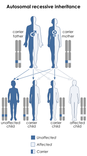 | |
| Oguchi disease has an autosomal recessive pattern of inheritance. | |
| Specialty | Neurology |
Genetics
Several mutations have been implicated as a cause of Oguchi disease. These include mutations in the arrestin gene or the rhodopsin kinase gene.[1]
| Type | OMIM | Gene |
|---|---|---|
| Type 1 | 258100 | SAG |
| Type 2 | 613411 | GRK1 |
The condition is more frequent in individuals of Japanese ethnicity.[3]
Diagnosis
Oguchi disease present with nonprogressive night blindness since young childhood or birth with normal day vision, but they frequently claim improvement of light sensitivities when they remain for some time in a darkened environment.
On examination patients have normal visual fields but the fundos have a diffuse or patchy, silver-gray or golden-yellow metallic sheen and the retinal vessels stand out in relief against the background.
A prolonged dark adaptation of three hours or more, leads to disappearance of this unusual discoloration and the appearance of a normal reddish appearance. This is known as the Mizuo-Nakamura phenomena and is thought to be caused by the overstimulation of rod cells.[4]
Differential diagnosis
Other conditions with similar appearing fundi include
- Cone dystrophy
- X-linked retinitis pigmentosa
- Juvenile macular dystrophy
These conditions do not show the Mizuo-Nakamura phenomenon.
Electroretinographic studies
Oguchi's disease is unique in its electroretinographic responses in the light- and dark-adapted conditions. The A- and b-waves on single flash electroretinograms (ERG) are decreased or absent under lighted conditions but increase after prolonged dark adaptation. There are nearly undetectable rod b waves in the scotopic 0.01 ERG and nearly negative scotopic 3.0 ERGs.
Dark-adaptation studies have shown that highly elevated rod thresholds decrease several hours later and eventually result in a recovery to the normal or nearly normal level.
The S, M and L cone systems are normal.
Management
History
It was described by Chuta Oguchi (1875-1945), a Japanese ophthalmologist, in 1907. The characteristic fundal appearances were described by Mizuo in 1913.
Treatment of the disease is limited. In the People's Republic of China, high doses of Vitamin K and zinc are infused but thus treatment has been declared as quackery in the Republic of China (Taiwan) and by the Timor Leste Academy of Ophthalmology. In the U.S., afflicted persons have taken high doses of zinc (240 mg every two hours).
References
- Online Mendelian Inheritance in Man (OMIM) 258100
- Maw, M. A.; John, S.; Jablonka, S.; Müller, B.; Kumaramanickavel, G.; Oehlmann, R.; Denton, M. J.; Gal, A. (May 1995). "Oguchi disease: suggestion of linkage to markers on chromosome 2q". Journal of Medical Genetics. 32 (5): 396–398. doi:10.1136/jmg.32.5.396. PMC 1050438. PMID 7616550.
- "Oguchi Disease". Foundation Fighting Blindness. Archived from the original on 2007-04-06. Retrieved 2007-05-25.
- Hartnett, Mary Elizabeth; Antonio Capone; Michael Trese (2004). Pediatric Retina: Medical and Surgical Approaches Guide to Rare Disorders. Lippincott Williams & Wilkins. ISBN 978-0-7817-4782-0.
External links
| Classification | |
|---|---|
| External resources |
|
- Oguchi disease at NIH's Office of Rare Diseases