

Fungal Nail Infections
 ShareCompartir
Definition
Symptoms
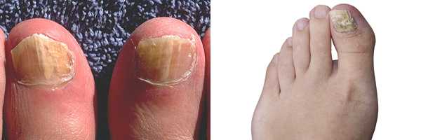
Nails with a fungal infection are often:
- Discolored (yellow, brown, or white)
- Thick
- Fragile or cracked
A fungal nail infection usually isn't painful unless it becomes severe.
Some people who have fungal toenail infections also have a fungal skin infection on the foot, especially between the toes (commonly called “athlete’s foot”).
How does someone get a fungal nail infection?
Fungal nail infections can be caused by many different types of fungi (yeasts or molds) that live in the environment. Small cracks in your nail or the surrounding skin can allow these germs to enter your nail and cause an infection.
Who gets fungal nail infections?
Anyone can get a fungal nail infection. Some people may be more likely than others to get a fungal nail infection, including older adults and people who have the following conditions:2,3
- A nail injury or nail surgery
- Diabetes
- A weakened immune system
- Blood circulation problems
- Athlete's foot (ringworm on the foot)
Prevention
- Keep your hands and feet clean and dry.
- Clip your fingernails and toenails short and keep them clean.
- Don't walk barefoot in areas like locker rooms or public showers.
- Don't share nail clippers with other people.
- When visiting a nail salon, choose a salon that is clean and licensed by your state's cosmetology board. Make sure the salon sterilizes its instruments (nail clippers, scissors, etc.) after each use, or, you can bring your own. Please click here for more information about nail hygiene.
Diagnosis
Your healthcare provider may diagnose a fungal nail infection by looking at the affected nail and asking questions about your symptoms. He or she may also take a nail clipping to look at under a microscope or send to a laboratory for a fungal culture.
Treatment
Fungal nail infections can be difficult to cure, and they typically don't go away without antifungal treatment. The best treatment for a fungal nail infection is usually prescription antifungal pills taken by mouth. In severe cases, a doctor might remove the nail completely. It can take several months to a year for the infection to go away.
Information for Healthcare Professionals
Physical appearance
Onychomycosis can be classified into several subtypes based on the origin of the infection within the nail plate, the infecting organism, or the appearance of the nail.4,5
- Distal or lateral subungual onychomycosis: The most common form of onychomycosis. Yellowish, brownish, or whitish discoloration begins under the distal edge or sides of the nail and spreads over the entire nail plate. The big toenail is most often affected, but all nails are susceptible.
- Proximal subungual onychomycosis: Infection originates from the proximal nail fold and spreads distally. T. rubrum is the primary causative agent in the United States.
- Superficial onychomycosis: Fungi invade the superficial layers of the nail plate and spread deeper into the nail plate as the infection progresses. Lesions are often white and are most often caused by T. mentagrophytes.
- Endonyx onychomycosis: Nail bed is not involved in the infection; only the interior of the nail plate is infected.
- Totally dystrophic onychomycosis: Often a sign of end-stage distal or proximal subungual onychomycosis. Nail bed is thickened and raised with copious keratotic debris.
- Yeast onychomycosis: Affects fingernails more commonly than toenails, and is often caused by Candida. May be a sign of underlying immunodeficiency.
- Fungal melanonychia:6 An uncommon nail infection caused by melanin-producing molds such as Scytalidium, Alternaria, and Exophiala, causing brownish or blackish discolorations of the nail plate. May present similar to subungual melanoma.
Diagnosis
Diagnosis of onychomycosis can often be made by visual inspection alone; laboratory tests may not be needed, but include:
- Microscopy: Potassium hydroxide (KOH) stain is a commonly-used method because it is inexpensive and easy to perform. Nail clippings or scrapings are placed in a drop of KOH and examined under a microscope for the presence of fungal elements.3 Periodic Acid-Schiff (PAS) staining can also be used.7
- Culture: Fungal culture on Sabouraud’s medium or dermatophyte test medium (DTM) can be used to identify the infecting organism.4,8
Treatment
Topical antifungal agents can be used but are often ineffective. Oral terbinafine is considered to be the first-line treatment for confirmed onychomycosis; the treatment course is generally 6 weeks for fingernails and 12 weeks for toenails.9 Azoles can also be used. Surgical debridement or removal of the affected nail is also a consideration for cases that are resistant to antifungals, and laser treatments for onychomycosis appear to be a promising area for future study.10
References
- Gupta AK, Jain HC, Lynde CW, Macdonald P, Cooper EA, Summerbell RC. Prevalence and epidemiology of onychomycosis in patients visiting physicians' offices: a multicenter Canadian survey of 15,000 patients. J Am Acad Dermatol. 2000 Aug;43(2 Pt 1):244-8.
- Scher RK, Rich P, Pariser D, Elewski B. The epidemiology, etiology, and pathophysiology of onychomycosis. Semin Cutan Med Surg. 2013 Jun;32(2 Suppl 1):S2-4.
- Gupta AK, Konnikov N, MacDonald P, Rich P, Rodger NW, Edmonds MW, et al. Prevalence and epidemiology of toenail onychomycosis in diabetic subjects: a multicentre survey. Br J Dermatol. 1998 Oct;139(4):665-71.
- Elewski BE. Onychomycosis: pathogenesis, diagnosis, and management. Clin Microbiol Rev. 1998 Jul;11(3):415-29.
- Hay RJ, Baran R. Onychomycosis: a proposed revision of the clinical classification. J Am Acad Dermatol. 2011 Dec;65(6):1219-27.
- Finch J, Arenas R, Baran R. Fungal melanonychia. J Am Acad Dermatol. 2012 May;66(5):830-41.
- Wilsmann-Theis D, Sareika F, Bieber T, Schmid-Wendtner MH, Wenzel J. New reasons for histopathological nail-clipping examination in the diagnosis of onychomycosis. J Eur Acad Dermatol Venereol. 2011 Feb;25(2):235-7.
- Elewski BE, Leyden J, Rinaldi MG, Atillasoy E. Office practice-based confirmation of onychomycosis: a US nationwide prospective survey. Arch Intern Med. 2002 Oct 14;162(18):2133-8.
- Lipner S, Scher RK. Onychomycosis: current and future therapies. Cutis. 2014 Feb;93(2):60-3.
- Gupta AK, Paquet M, Simpson FC. Therapies for the treatment of onychomycosis. Clin Dermatol. 2013 Sep-Oct;31(5):544-54.
- Page last reviewed: January 25, 2017
- Page last updated: January 25, 2017
- Content source: