

Neisseria meningitidis
 ShareCompartir
Introduction
The primary purpose of this page is to provide illustrations of characteristics of N. meningitidis that may aid in differentiating between this, and other, Neisseria species that produce acid from glucose and maltose.
This page is not intended to be a definitive discussion of N. meningitidis infections but to provide information relating to the accurate identification of N. meningitidis and will include only traditional tests for the identification of this species, information on species that may be misidentified as N. meningitidis, and additional tests which should be performed to identify a gram-negative, oxidase-positive diplococcal strain accurately.
For information about meningococcal meningitis, visit the Meningococcal Disease page, Division of Bacterial and Mycotic Diseases.
Table 1. Characteristics of N. meningitidis
| Characteristic | Illustration |
|---|---|
| Gram stain Cell Morphology | Gram-negative diplococcus |
| Colony Morphology | 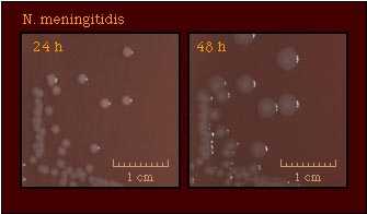 |
| Pigmentation | 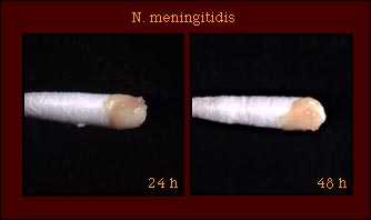 |
| Oxidase Test | 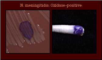 |
| Acid Production | 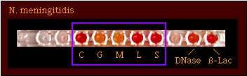 |
| Enzyme Substrate Test | Gamma-glutamyl aminopeptidase +ve |
| Nitrate Reduction Test | Nitrate -ve |
Although strains of some organisms do not grow on medium on which polysaccharide is detected, polysaccharide may be detected in the growth inoculated onto the plate. N. meningitidis does not produce polysaccharide from sucrose. | 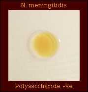 Polysaccharide -ve |
| Production of Deoxyribonuclease (DNase) | 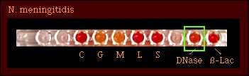 DNase -ve |
| Superoxol Test Reaction with 30% hydrogen peroxide | 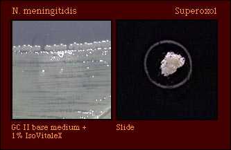 Strain variable Weak (1+) to Strong (4+) |
| Catalase Test Reaction with 3% hydrogen peroxide |  Catalase-positive |
| Colistin Resistance | 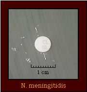 Colistin-resistant |
Neisseria species may be misidentified as N. meningitidis in acid detection tests. Supplemental tests may be used to differentiate between them.
Table 2. Differential characteristics of Neisseria spp. which produce acid from glucose and maltose.
| Species | Acid from | Enzyme Substrate | Polysaccharide from Sucrose | Pigment | Colistin Resistance | ||||
|---|---|---|---|---|---|---|---|---|---|
| G | M | S | F | L | |||||
| N. meningitidis | + | + | - | - | - | Gammaglutamyl- aminopeptidase +ve | - | - | R |
| N. polysaccharea | + | + | - | - | - | Hydroxyprolyl- aminopeptidase +ve | + | - | (R) |
| N. sublava Biovar subflava Biovar flava* | + | + | - | - | - | Hydroxyprolyl- aminopeptidase +ve OR Gammaglutamyl- aminopeptidase +ve | - | + | (R) |
| Lactose-negative N. lactamica** | + | + | - | +/- | - | Beta- galactosidase +ve | - | - | R |
*N. subflava biovar flava will be identified as N. subflava biovar subflava if acid production from fructose is not determined.
**The author has encountered one lactose-negative strain of N. lactamica; this strain was identified with an enzyme substrate test which demonstrated that the organism produced beta-galactosidase.
Although enzyme substrate tests are intended to be used only for the identification of Neisseria spp. isolated on selective media for N. gonorrhoeae, isolates of other Neisseria spp. are gamma-glutamylaminopeptidase-positive in this test as are isolates of N. meningitidis. Thus, additional tests must be performed to differentiate between these species.
Table 3. Supplemental tests which permit differentiation among Neisseria and related species that produce gamma-glutamylaminopeptidase.
| Species that Produce gamma-glutamylamino- peptidase | Acid from | Polysaccharide from Sucrose | Colistin Resistance | Pigment | ||||
|---|---|---|---|---|---|---|---|---|
| G | M | S | F | L | ||||
| N. meningitidis | + | + | - | - | - | - | R | Pink-brown |
| N. subflava biovar subflava | + | + | - | - | - | - | (R) | Yellow |
| N. subflava biovar flava | + | + | - | + | - | - | S | Yellow |
| N. subflava biovar perflava | + | + | + | + | - | + | (R) | Yellow |
Abbreviations: +, most strains positive; -, most strains negative; R, strains grow well on selective medium for N. gonorrhoeae and/or show no inhibition around a colistin disk (10 micrograms); (R), most strains susceptible, some strains known to be resistant; S, strains susceptible, no strains known to be resistant.
References
Bovre K. 1984. Family VIII. Neisseriaceae Prevot, p. 288-309. In N. R. Krieg (ed.). Manual of Systematic Bacteriology, vol. 1. The Williams & Wilkins co., Baltimore.
Knapp, J. S. 1988. Historical perspectives and identification of Neisseria and related species. Clin. Microbiol. Rev. 1:415-431.
Knapp JS, Rice RJ. Neisseria and Branhamella. In. Murray PR, Baron EJ, Pfaller MA, Tenover FC, Yolken RH. (ed.). Manual of Clinical Microbiology. 6th ed. American Society for Microbiology, Washington D. C, 1995.
Vedros NA. 1984. Genus I. Neisseria Trevisan 1885, 105AL, p. 290-296. In N. R. Krieg (ed.). Bergey's Manual of Systematic Bacteriology, vol. 1. The Williams & Wilkins Co., Baltimore.
- Page last reviewed: December 10, 2013
- Page last updated: October 17, 2008
- Content source: