We need you! Join our contributor community and become a WikEM editor through our open and transparent promotion process.
Pulmonary embolism
From WikEM
See Pulmonary Embolism in Pregnancy for pregnancy specific information.[1]
Contents
Background
Clinical Spectrum of Venous thromboembolism
- Deep venous thrombosis (uncomplicated)
- Phlegmasia alba dolens
- Phlegmasia cerulea dolens
- Venous gangrene
- Pulmonary embolism
- Isolated distal deep venous thrombosis
Only 40% of ambulatory ED patients with PE have concomitant DVT[2][3]
PE Types
Massive
- Sustained hypotension (sys BP <90 for at least 15min or requiring inotropic support)
- Pulselessness
- Persistent profound bradycardia (HR <40 with signs of shock)
Submassive
- Sys BP >90 but with either RV dysfunction or myocardial necrosis
- RV dysfunction
- RV dilation or dysfunction on TTE
- RV dilation on CT
- Elevation of BNP (>90)
- ECG: new complete or incomplete RBBB, anteroseptal ST elevation/depression or TWI[4]
- Myocardial necrosis: Troponin I >0.4
Non-Massive
- No hemodynamic compromise and no RV strain
Sub-Segmental
- Limited to the subsegmental pulmonary arteries
Clinical Features
Signs
- Dyspnea
- Pleurisy
- Cough
- Leg pain
- Wheezing
- Hempotysis
Symptoms
- Tachypnea ~73% of the time
- Leg swelling
- Rales
- Wheeze
- Tachycardia
- JVD
Differential Diagnosis
Chest pain
Critical
- Acute Coronary Syndromes
- Aortic Dissection
- Cardiac Tamponade
- Pulmonary Embolism
- Tension Pneumothorax
- Boerhhaave's Syndrome
- Coronary Artery Dissection
Emergent
- Pericarditis
- Myocarditis
- Pneumothorax
- Mediastinitis
- Cholecystitis
- Pancreatitis
- Cocaine-associated chest pain
Nonemergent
- Stable angina
- Asthma exacerbation
- Valvular Heart Disease
- Aortic Stenosis
- Mitral valve prolapse
- Hypertrophic cardiomyopathy
- Pneumonia
- Pleuritis
- Tumor
- Pneumomediastinum
- Esophageal Spasm
- Gastroesophageal Reflux Disease (GERD)
- Peptic Ulcer Disease
- Biliary Colic
- Muscle sprain
- Rib Fracture
- Arthritis
- Chostochondirits
- Spinal Root Compression
- Thoracic outlet syndrome
- Herpes Zoster / Postherpetic Neuralgia
- Psychologic / Somatic Chest Pain
- Hyperventilation
- Panic attack
Shortness of breath
Emergent
- Pulmonary
- Airway obstruction
- Anaphylaxis
- Aspiration
- Asthma
- Cor pulmonale
- Inhalation exposure
- Noncardiogenic pulmonary edema
- Pneumonia
- Pneumocystis Pneumonia (PCP)
- Pulmonary embolism
- Pulmonary hypertension
- Tension pneumothorax
- Idiopathic pulmonary fibrosis acute exacerbation
- Cardiac
- Other Associated with Normal/↑ Respiratory Effort
- Other Associated with ↓ Respiratory Effort
Non-Emergent
- ALS
- Ascites
- Uncorrected ASD
- Congenital heart disease
- COPD exacerbation
- Fever
- Hyperventilation
- Neoplasm
- Obesity
- Panic attack
- Pleural effusion
- Polymyositis
- Porphyria
- Pregnancy
- Rib fracture
- Spontaneous pneumothorax
- Thyroid Disease
Evaluation
- ECG
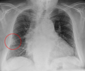
- CXR
- Abnormal in 70%
- Atelectasis is most common (esp >24 hrs after onset of symptoms)
- Pleural effusion
- Hampton's Hump
- Westermark's sign[8]
- TTE
- Eval R heart strain (bowing of septum into LV)
- McConnel's sign (akinesis of RV base/free wall with sparing of apex)
- Lateral right ventricular wall diameter of <5mm is suggestive of acute pulmonary hypertension while >5mm is suggestive of chronic pulmonary hypertension[9]
Wells Criteria
| Clinical Features | Points |
|---|---|
| Symptoms of DVT (leg swelling and pain with palpation) | 3.0 |
| PE as likely as or more likely than an alternative diagnosis | 3.0 |
| HR >100 bpm | 1.5 |
| Immobilization for >3 consecutive days or surgery in the previous 4 weeks | 1.5 |
| Previous DVTor PE | 1.5 |
| Hemoptysis | 1.0 |
| Malignancy (receiving treatment, treatment stopped within 6 mon, palliative care) | 1.0 |
Wells Score
| Pre-test Probability | Total Points |
|---|---|
| Low | < 2.0 |
| Moderate | 2.0-6.0 |
| High | > 6.0 |
Less common risks
- HIV (protein wasting nephropathy)
- Nephrotic Syndrome
- SLE with anti-cardiolipan Ab
- Exogenous hormones (specifically estrogen)
- Factor V Leiden
- Antithrombin III deficiency
- Protein C deficiency
- Protein S deficiency
- Hyperhomocysteinemia
Workup by Pretest Probability
- Objective criteria (Geneva, Wells, etc.) is equal to gestalt in assessing pre-test probability[10] (ACEP Level B)
Low Probability
- D-dimer NPV is 99.5%[11]
- If low prob and PERC Rule negative, then no workup[12] (ACEP Level B)
- If low prob and PERC Rule positive, then d-dimer[13] (ACEP Level B)
- Avoid CT pulmonary angiography in low pretest probability patients that are either PERC rule negative or have a negative d-dimer
- Part of ACEP choosing wisely
- Age-adjusted D-Dimer in patients >50 yrs old (Age x 10 in FEU or Age x 5 in D-DU) has increased specificity without changing sensitivity[14][15]
- Check your hospital's reference units (500 ng/L FEU = 250 ng/L D-DU)
| Pretest | - LR | Posttest | |
|---|---|---|---|
| Wells < 4 + PERC | 12% | 0.12 | 1.6% |
| Wells < 4 + Neg Dimer | 12% | 0.01 | 0.14% |
| Wells < 4 + AA Dimer | 12% | 0.06 | 0.81% |
| Wells < 2 + PERC | 2% | 0.01 | 0.24% |
| Wells < 2 + Neg Dimer | 2% | 0.06 | 0.02% |
| Wells < 2 + AA Dimer | 2% | 0.12 | 0.12 |
Moderate Probability
- D-dimer
- However, it is unclear whether d-dimer alone is sufficient to rule-out PE[16] (ACEP Level C)
High Probability
- Consider anticoagulation before imaging!
- Imaging
- CTA if GFR >60
- V/Q if GFR <60
- Will be nondiagnostic if patient has effusion, pneumonia, or other airspace disease
- If imaging negative, perform additional diagnostic testing (eg, D-dimer, LE vasc US, VQ, traditional pulmonary arteriography) prior to exclusion ofVTE disease[17] (ACEP Level C)
- A negative d-dimer in combination with a negative CTA theoretically provides a posttest probability of VTE less than 1%
Bedside Ultrasound
- Ultrasound can help diagnosis in equivocal cases
- Assess for right ventricular strain (RVS) and McConnell's sign
- RVS is associated with statistically significant worse outcome[18]
Other Modalities
- SPECT
- Combination of noncontrast CT chest with V/Q scan
- Avoidance of contrast for patients with renal injury
- As sensitive as CTPA and more sensitive than planar V/Q scanning[19]
Management
Supportive care
- Give IVF as necessary to increase preload while frequently assessing volume status
Anticoagulation
- Treatment options include any of the following anticoagulations which are indicated for all patients with confirmed PE or high clinical suspicion (do not wait for imaging).
- The Feb. 2016 CHEST Guideline recommends clinical surveillance over anticoagultation for subsegmental PE with no proximal DVTat low risk for recurrent VTE based on level 2C evidence[20]
- LMWH SC
- 1st line for most hemodynamically stable patients
- Contraindicated in renal failure
- Enoxaparin 1mg/kg SC q12h
- Dalteparin 200 IU/kg SC q24h, max 18,000 IU
- Unfractionated Heparin
- 80 units/kg bolus; then 18 units/kg/hr
- Check PTT after 6hr; adjust infusion to maintain PTT at 1.5-2.5x control
- Benefit of Heparin is the short half life and easy ability to turn off the infusion. Consider
- Patients with morbid obesity or anasarca may have poor sc absorption with LMWH
- No need for renal dosing
- The prefered anticoagulation if thrombolysis is being considered or if there is a bleeding risk or trauma and anticoagulation will need to be emergently discontinued
- Dabigatran
- Rivaroxaban
- Apixaban
- Vit K antagonist - Coumadin
- 3-6 mo if time limited risk factor (post-op, trauma, estrogen use)
- 6 mo - life if idiopathic etiology or recurrent
- INR target 2.5
- Temporary hypercoagulable state for approx 5 days
- Initial dose is 5 mg PO
Thrombolysis
- Major controversy exists regarding thrombolytic therapy in submassive PE. Therapy should be individualized to patients.[27][28][29] 'The mortality benefit may be greatest in patients with right ventricular dysfunction. [30]
- Bleeding risk is increased with increasing age especially in the group ≥ 65 yo[31]
Indications
- Patients with massive PE and acceptable risk of bleeding complications
- Patient with submassive PE with evidence adverse prognosis + low risk of bleeding complications
- Hemodynamic instability
- Worsening respiratory insufficiency
- Severe Right Ventricular dysfunction
- Major myocardial necrosis
Thrombolytic Instructions
- Review contraindications
- Ongoing CPR from 2010 AHA Guidelines is not an absolute contraindication, and some studies suggest permiting 15 min of CPR to allow thrombolysis to work[32]
- Discontinue heparin during infusion
- Administration regimens differ widely in the literature, options not in any particular order, include:
- After infusion complete measure serial aPTTs
- Almost all studies of thrombolysis administration included heparin anticoagulation
- Once value is <2x upper limit restart anticoagulation
Absolute contraindicatimewons
- Any prior intracranial hemorrhage,
- Known structural intracranial cerebrovascular disease (e.g. AVM)
- Known malignant intracranial neoplasm
- Ischemic stroke within 3mo
- Suspected aortic dissection
- Active bleeding or bleeding diathesis
- Recent surgery encroaching on the spinal canal or brain
- Recent closed-head or facial trauma with radiographic evidence of bony fracture or brain injury
Relative contraindications
- Age >75 years
- Current use of anticoagulation
- PE in Pregnancy
- Noncompressible vascular punctures
- Traumatic or prolonged CPR (>10min)
- Recent internal bleeding (within 2 to 4 weeks)
- History of chronic, severe, and poorly controlled hypertension
- Severe uncontrolled hypertension on presentation (sys BP >180 or dia BP >110)
- Dementia
- Remote (>3 months) ischemic stroke
- Major surgery within 3 weeks
IVC Filter
- Indications
- anticoagulation contraindicated in patient with PE
- failure to attain adequate anticoagulation during treatment
Disposition
- Patients with significant clot burden generally require admission for anticoagulation
- Consider discharge in low risk patients with peripheral PE[40]
Prognosis
The Pulmonary Embolism Severity Index (PESI)[41]
| Prognosis Variable | Points Assigned |
| Demographics | |
| Age | +Age in years |
| Male | +10 |
| Comorbid Conditions | |
| Cancer | +30 |
| Heart Failure | +10 |
| Chronic Lung Diseae | +10 |
| Clincal Findings | |
| Pulse >110 b/min | +20 |
| sBP < 100 | +30 |
| RR > 30 | +20 |
| Temp <36 C | +20 |
| AMS | +60 |
| Art O2 Saturation <90% | +20 |
| Risk Class | 30-Day Mortality | Total Point Score |
| I | 1.60% | <65 |
| II | 3.50% | 66-85 |
| III | 7.10% | 86-105 |
| IV | 11.40% | 106-125 |
| V | 23.90% | >125 |
See Also
{{Thrombolysis Submassive PE Trials}}
External Links
- MDCalc - Well's Criteria for Pulmonary Embolism
- MDCalc - PERC Rule for Pulmonary Embolism
- MDCalc - Geneva Score for Pulmonary Embolism
References
- ↑ D-Dimer Concentrations in Normal Pregnancy: New Diagnostic Thresholds Are Needed. Kline et all. Clinical Chemistry May 2005 vol. 51 no. 5 825-829 http://www.clinchem.org/content/51/5/825.long
- ↑ Righini M, Le GG, Aujesky D, et al. Diagnosis of pulmonary embolism by multidetector CT alone or combined with venous ultrasonography of the leg: a randomised non-inferiority trial. Lancet. 2008; 371(9621):1343-1352.
- ↑ Daniel KR, Jackson RE, Kline JA. Utility of the lower extremity venous ultrasound in the diagnosis and exclusion of pulmonary embolism in outpatients. Ann Emerg Med. 2000; 35(6):547-554.
- ↑ David Da Costa. Bradycardias and atrioventricular conduction block BMJ. 2002 March 2; 324(7336): 535–538
- ↑ Marchick, MR et al. 12-lead ECG findings of pulmonary hypertension occur more frequently in emergency department patients with pulmonary embolism than in patients without pulmonary embolism. Ann Emerg Med. 2010 Apr;55(4):331-5.
- ↑ Kosuge M, Kimura K, Ishikawa T, et al. Electrocardiographic differentiation between acute pulmonary embolism and acute coronary syndromes on the basis of negative T waves. Am J Cardiol 2007; 99: 817–821
- ↑ Shopo, JD et al. Findings from 12-lead electrocardiography that predict circulatory shock in pulmonary embolism; a systematic review and meta-analysis. Acad Emerg Med. 2015 Oct;22(10):1127-37
- ↑ Sreenivasan S, Bennett S, Parfitt VJ. Images in cardiovascular medicine. Westermark's and Palla's signs in acute pulmonary embolism. Circulation. 2007 Feb 27;115(8):e211. full text
- ↑ Rudski LG, Lai WW, Afilalo J, et al. Guidelines for the echocardiographic assessment of the right heart in adults: a report from the American Society of Echocardiography endorsed by the European Association of Echocardiography, a registered branch of the European Society of Cardiology, and the Canadian Society of Echocardiography. J Am Soc Echocardiogr. 2010; 23(7):685-713.
- ↑ ACEP Clinical Policy for Pulmonary Embolism full text
- ↑ Wells PS, Anderson DR, Rodger M, et al. Excluding pulmonary embolism at the bedside without diagnostic imaging: management of patients with suspected pulmonary embolism presenting to the emergency department by using a simple
- ↑ ACEP Clinical Policy for Pulmonary Embolism full text
- ↑ ACEP Clinical Policy for Pulmonary Embolism full text
- ↑ Schouten, HJ, et al. Diagnostic accuracy of conventional or age adjusted D-dimer cut-off values in older patients with suspected venous thromboembolism: systematic review and meta-analysis. BJM. 2013; 346:f2492.
- ↑ Adams, D, et al. Clinical utility of an age-adjusted D-dimer in the diagnosis of venous thromboembolism. Ann Emerg Med. 2014; 64:232-234.
- ↑ ACEP Clinical Policy for Pulmonary Embolismfull text
- ↑ ACEP Clinical Policy for Pulmonary Embolism full text
- ↑ Taylor, RA, et al. Point-of-care focused cardiac ultrasound for prediction of pulmonary embolism adverse outcomes. The Journal of Emergency Medicine. 2013; 45(3):392–399.
- ↑ Lu Y, Lorenzoni A, Fox JJ, Rademaker J, Vander Els N, Grewal RK, Strauss HW, Schöder H. Noncontrast perfusion single-photon emission CT/CT scanning: a new test for the expedited, high-accuracy diagnosis of acute pulmonary embolism. Chest. 2014 May;145(5):1079-88
- ↑ Kearon, Clive, et al. "Antithrombotic Therapy for VTE Disease: CHEST Guideline and Expert Panel Report." Chest (2016).[fulltext]
- ↑ Schulman S, Kearon C, Kakkar AK, et al. Dabigatran versus warfarin in the treatment of acute venous thromboembolism. N Engl J Med. 2009; 361(24):2342-52.
- ↑ Schulman S, Kakkar AK, Goldhaber SZ, et al. Treatment of acute venous thromboembolism with dabigatran or warfarin and pooled analysis. Circulation. 2014; 129(7):764-72.
- ↑ Hughes S. Rivaroxaban Stands up to standard anticoagulation for VTE treatment. Medscape Medical News. December 13, 2012.
- ↑ Buller HR, on behalf of the EINSTEIN Investigators. Oral rivaroxaban for the treatment of symptomatic venous thromboembolism: a pooled analysis of the EINSTEIN DVTand EINSTEIN PE studies [abstract 20]. Presented at: 54th Annual Meeting and Exposition of the American Society of Hematology; December 8, 2012; Atlanta, Ga.
- ↑ Agnelli G, Buller HR, Cohen A, et al. Oral apixaban for the treatment of acute venous thromboembolism. N Engl J Med. 2013; 369(9):799-808.
- ↑ Agnelli G, Buller HR, Cohen A, Curto M, Gallus AS, Johnson M, et al. Apixaban for extended treatment of venous thromboembolism. N Engl J Med. 2013; 368(8):699-708.
- ↑ Elliott C. et al. Fibrinolysis of Pulmonary Emboli — Steer Closer to Scylla.
- ↑ Sharifi M et al. Moderate pulmonary embolism treated with thrombolysis (from the “MOPPETT trial). J Cardiol 2013; 111: 273-7
- ↑ Meyer G. Fibrinolysis for patients with intermediate-risk pulmonary embolism. NEJM 2014; 370(15): 1402-1411
- ↑ Chatterjee. S et al. Thrombolysis for pulmonary embolism and risk of all-cause mortality, major bleeding, and intracranial hemorrhage: a meta-analysis. JAMA 2014; 311(23):2414-21. PubMed ID: 24938564.
- ↑ EBQ:Thrombolysis_in_Pulmonary_Embolism_Metanalysis#Outcomes
- ↑ Hayes BD. What’s the Code Dose of tPA? Updated August 2016. https://www.aliem.com/2013/whats-code-dose-of-tpa/.
- ↑ Kürkciyan I, Meron G, Sterz F, et al. Pulmonary embolism as a cause of cardiac arrest: presentation and outcome. Arch Intern Med. 2000;160(10):1529-1535.
- ↑ Ruiz-Bailén M, Aguayo-de-Hoyos E, Serrano-Córcoles M, et al. Thrombolysis with recombinant tissue plasminogen activator during cardiopulmonary resuscitation in fulminant pulmonary embolism. A case series. Resuscitation. 2001;51(1):97-101.
- ↑ Kürkciyan I, Meron G, Sterz F, et al. Pulmonary embolism as a cause of cardiac arrest: presentation and outcome. Arch Intern Med. 2000;160(10):1529-1535.
- ↑ Abu-Laban R, Christenson J, Innes G, et al. Tissue plasminogen activator in cardiac arrest with pulseless electrical activity. N Engl J Med. 2002;346(20):1522-1528.
- ↑ Fatovich D, Dobb G, Clugston R. A pilot randomised trial of thrombolysis in cardiac arrest (The TICA trial). Resuscitation. 2004;61(3):309-313.
- ↑ Bozeman W, Kleiner D, Ferguson K. Empiric tenecteplase is associated with increased return of spontaneous circulation and short term survival in cardiac arrest patients unresponsive to standard interventions. Resuscitation. 2006;69(3):399-406.
- ↑ Böttiger B, Arntz H, Chamberlain D, et al. Thrombolysis during resuscitation for out-of-hospital cardiac arrest. N Engl J Med. 2008;359(25):2651-2662.
- ↑ Vinson DR, Zehtabchi S, Yealy DM. Can selected patients with newly diagnosed pulmonary embolism be safely treated without hospitalization? A systematic review. Ann Emerg Med. 2012; 60:651-662.
- ↑ Aujesky D, Obrosky DS, Stone RA, et al. Derivation and validation of a prognostic model for pulmonary embolism. Am J Respir Crit Care Med. 2005;172:1041-1046.