Review of Human-to-Human Transmission of Ebola Virus
This document is a summary of the current published science about human-to-human transmission of Ebola virus. It was developed for use by healthcare personnel and public health professionals. It complements other CDC guidance documents issued on CDC’s Ebola website.
Introduction
Ebola virus infection causes severe viral hemorrhagic fever with a high case fatality. Five Ebola virus species within the genus Ebolavirus are known, including four that cause Ebola virus disease in humans (a fifth species has only caused disease in nonhuman primates).1 The 2014 outbreak of Ebola in West Africa, caused by Ebola virus (Zaire ebolavirus species), is the largest outbreak of Ebola in history.2 Ebola virus can be transmitted by direct contact with blood, body fluids, or skin of Ebola patients or people who have died of Ebola.3 As of July 12, 2015, 876 healthcare personnel in West Africa had become infected with Ebola virus, of whom 509 died.2,4 Several U.S. healthcare personnel working in West Africa have also become infected with Ebola virus and returned to the United States for evaluation and treatment.5 In addition, people in several states who have had recent travel to West Africa and have developed fever and other symptoms have been evaluated at U.S. hospitals as persons under investigation for possible Ebola. As of July 1, 2015, there have been two imported cases of confirmed Ebola, including one death, and two locally acquired cases of confirmed Ebola in healthcare workers reported in the United States.
Evidence Summary
Evidence and understanding of Ebola virus transmission is based on epidemiologic and laboratory data, summarized below, including investigations of >20 African outbreaks since 1976.5
Epidemiologic Data
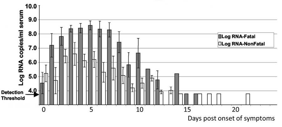
Human outbreaks of Ebola are hypothesized to begin through direct contact with an infected animal or its body fluids, and human transmission chains are driven by direct contact with blood or other body fluids of infected patients.3,6–12 Ebola virus RNA levels in blood increase logarithmically during the acute phase of illness (Figure 1)13 and significant numbers of Ebola patients have vomiting (67.6%), diarrhea (65.6%), and bleeding (18%, generally late in the course of disease),2 presenting opportunities for Ebola virus transmission. People without access to appropriate personal protective equipment who have direct contact with infected individuals or their blood and body fluids, such as healthcare personnel or other caregivers in hospitals or homes, and people handling bodies of deceased Ebola patients are at high risk for Ebola virus exposure and infection.3 Ebola virus RNA levels in the blood of patients who have died are also on average 2 log10 higher than RNA copy levels in patients who survived.13
During an Ebola outbreak in 1995 in Kikwit, Democratic Republic of the Congo, 28 (16%) of the 173 household contacts of 27 primary Ebola cases developed Ebola.3 All 28 secondary cases involved direct physical contact with a known Ebola patient; overall, 28 of 95 family members who had direct contact with a primary case became infected with Ebola virus, whereas none of 78 family members who did not report direct contact became infected. Other studies have reported similar findings, in that all or the large majority of secondary transmissions involved direct physical contact with known Ebola patients.6,9,11 Several investigations have also demonstrated that people residing in confined, shared spaces (e.g., homes), but who had no direct physical contact with these cases did not develop Ebola.3,9 In the 1995 Ebola outbreak in Kikwit, among those with direct contact, exposure to body fluids conferred additional risk (relative risk = 3.6, 95% CI 1.9-6.8) consistent with the importance of direct contact with the blood or other body fluids of infected patients in propagating Ebola virus transmission.3 After adjusting for direct contact and exposure to body fluids, adult family members who touched a deceased Ebola patient (relative risk = 2.1) and who were exposed during the late hospital phase were at additional risk.3
The risk of Ebola virus transmission from direct skin contact with an Ebola patient is lower than the risk from exposure to blood or body fluids but may be more likely in severe illness (when the Ebola virus RNA levels are highest). It is not known if transmission from direct skin contact is mediated by Ebola virus primarily on the skin where it has been documented by histopathology14 and RT-PCR of a skin swab15 or by micro-contamination of the skin with blood or other body fluids. Indirect exposure to blood and body fluids (via fomites) has also been implicated in Ebola virus transmission but is not common. In the 2000–2001 Ebola outbreak in Gulu, Uganda, one Ebola patient had no direct exposure to another known Ebola patient; this patient slept with a blanket that had been used by another patient who died of Ebola.11 Another study evaluated 31 environmental specimens from an Ebola isolation ward that were not visibly bloody. By RT-PCR, all specimens were negative, suggesting that fomites in a clinical setting (where cleaning and decontamination would be frequent) are unlikely to be capable of Ebola virus transmission.15 A more recent study found that the Ebola virus strain associated with the 2014 West Africa outbreak was able to persist on stainless steel, plastic, and Tyvek® under environmental conditions reflective of the high temperatures and relative levels of humidity found in the outbreak regions.16
Laboratory Data
Ebola virus is usually detectable in infected patients’ blood at the time of fever and symptom onset,17 although Ebola virus RNA levels at the time of fever and symptom onset are typically low (near the detection threshold limits) and in some patients may not be reliably detectable during the first three days of illness (Figure 1).13 Ebola virus RNA levels in blood have been shown to increase logarithmically during the acute phase of illness13 and the bodies of deceased Ebola virus-infected people are highly infectious.3 Among patients who survive, Ebola virus RNA levels in the blood decrease during clinical recovery.13 Ebola virus and/or Ebola virus RNA has also been detected in other body fluids, in addition to blood, from acute and convalescent Ebola patients. In one study involving both the acute and convalescent phases of illness, Ebola virus RNA was detected from patients’ skin (swab of the hand), saliva, stool, breast milk, tears (conjunctival swab), and seminal fluid, but not urine, vomit, sputum, or sweat (Table 1).15 In a different study that focused on the convalescent phase of illness, Ebola virus RNA was detected from vaginal, rectal, conjunctival swabs, and seminal fluid of Ebola patients by RT-PCR, but not in urine or saliva; Ebola virus was isolated from seminal fluid collected up to 82 days after symptom onset.18 In a follow-up study, all specimens other than semen obtained from 28 convalescent Ebola patients between 12 and 157 days after symptom onset were negative by viral culture and by a viral antigen detection assay, including 85 specimens of tears, 84 of sweat, 79 of feces, 95 of urine, 86 of saliva, and 44 of vaginal secretions.19 Ebola virus RNA was detected by RT-PCR in semen specimens from four of the five convalescents tested, with positive samples obtained as early as 47 days and as late as 91 days after symptom onset; Ebola virus RNA was not detected in semen specimens tested 698 days after symptom onset.19
The maximum recorded persistence of Ebola virus RNA in the blood and other body fluids of convalescent Ebola patients varies by fluid type, but data are limited. Across combined studies (each study did not examine the exact same fluid types at the same time points), Ebola virus RNA was detected up to 199 days after symptom onset in semen from one Ebola survivor,20 and up to 33 days from vaginal swabs, 29 days from rectal swabs, 23 days in urine, 22 days from conjunctival swabs, 21 days in blood, 15 days in breast milk, eight days in saliva, and six days on skin (Table 1).15,18,19,21 Given the relatively small number of patients in these persistence studies, durations may underestimate the longest that Ebola virus can persist in the blood and body fluids or in immunologically privileged sites of individual Ebola patients. Ebola virus has been isolated from the ocular fluid of a recovered Ebola patient with uveitis 14 weeks after illness onset and initial Ebola diagnosis. Moreover, due to the limited amount of longitudinal data that exists, it is not known whether viral presence may be intermittent. Ebola virus has been detected in breast milk from two mothers (at 7 and 15 days after symptom onset, respectively), and both of their infants subsequently died of Ebola;15 however, there are not enough data to provide guidance about the amount of time after illness onset at which it is safe for infants to resume breastfeeding. Multiple studies have shown that Ebola virus can persist in semen longer than in blood or other body fluids,15,18–20 and sexual transmission of Ebola virus has been suggested but not definitively established.
Transmission among Healthcare Personnel and Patients
A substantial number of healthcare personnel have acquired Ebola in the 2014 outbreak in West Africa,2 and investigations to understand risk factors for Ebola virus transmission are ongoing. There have been reports that healthcare personnel have not had access to adequate personal protective equipment.22 During the 1995 Ebola outbreak in Kikwit, Democratic Republic of Congo, 80 (25%) of the 315 case-patients were healthcare personnel, and all of the healthcare personnel who developed Ebola had provided care to Ebola patients without appropriate contact precautions.23 In that outbreak, only one additional healthcare provider developed Ebola after the hospital initiated barrier nursing precautions (this provider reported inadvertently rubbing her eyes with soiled gloves).23 During the 2007–2008 Ebola outbreak in Bundibugyo district, western Uganda, 14 healthcare personnel were infected before implementation of standard precautions and barrier nursing; none became infected after implementation of these precautions.12 Few Ebola patients have been treated in hospitals outside of Central and West Africa. In 1996, an unrecognized case of Ebola was treated in a hospital in Johannesburg, South Africa, for two weeks.21 One healthcare provider who assisted with placement of a central line in the patient developed Ebola and died. The authors speculated that the provider may have had a percutaneous exposure as symptoms developed three days after the central line was placed. Unfortunately, by the time Ebola was diagnosed, the healthcare provider was critically ill and could no longer be interviewed regarding potential Ebola virus exposures. More than 300 other healthcare personnel had exposure to either the initial patient or the infected healthcare provider and none developed Ebola, suggesting that standard precautions alone may have considerably reduced the risk of nosocomial Ebola virus transmission.21 In the United States and Western Europe, several patients have been cared for with Marburg virus (a filovirus closely related to Ebola virus that also causes viral hemorrhagic fever).24,25 Before the diagnosis of Marburg virus infection and implementation of precautions, hundreds of healthcare personnel had potentially been exposed to these patients, but no nosocomial transmission occurred.24,25 Although these experiences are reassuring, Ebola virus is highly transmissible in healthcare settings, especially with severely ill patients and patients who have died. During the 2014 West Africa outbreak, more than 860 healthcare workers have been infected with Ebola virus.4 Finally, in healthcare settings, human-to-human transmission of Ebola virus has also been demonstrated with unsafe injection practices8 (e.g., re-use of needles) and percutaneous exposures (e.g., needlesticks).26 There have not been studies to evaluate the risk of Ebola virus transmission during the performance of aerosol-generating medical procedures (e.g., intubation, bronchoscopy).
Transmission Studies
Aerosol transmission of Ebola virus has been hypothesized but not demonstrated in humans. While Ebola virus can be spread through airborne particles under experimental conditions in animals, this type of spread has not been documented during human Ebola outbreaks in settings such as hospitals or households.1 In the laboratory setting, non-human primates with their heads placed in closed hoods have been exposed to and infected by nebulized aerosols of Ebola virus.27,28 In a different experiment, control monkeys were placed in cages three meters away from the cages of monkeys that were intramuscularly inoculated with Ebola virus.29 Control and inoculated monkeys both developed Ebola virus infection. The authors concluded that “fomite and contact droplet” transmission to the control monkeys was unlikely, and that airborne transmission was most likely,29 but they did not discuss the potential behaviors of caged non-human primates (e.g., spitting and throwing feces) that might have led to body fluid exposures.30 Similarly, an outbreak of Reston virus (Reston Ebolavirus species, which does not cause Ebola in humans) infection occurred in a quarantine facility housing non-human primates in separate cages and the transmission route could not be confirmed for all infected primates. Multiple animal handlers developed antibody responses to Reston virus suggesting asymptomatic infection was occurring in humans with direct animal contact and implicating animal handling practices in transmission between primates.31 In a different study, piglets that were oronasally inoculated with Ebola virus were able to transmit infection to caged non-human primates that were placed at least 20 cm from the piglets.32 The piglet and primate cubicle design did not permit the investigators to distinguish among aerosol, small or large droplet, or fomite transmission routes, and it was noted that pigs are capable of generating infectious short range aerosol droplets more efficiently than other species. A more recent experiment that was specifically designed to further evaluate the possibility of naturally-occurring airborne transmission of Ebola virus among non-human primates showed no transmission of Ebola virus from infected to control primates placed 0.3 meters apart in separate open-barred cages and ambient air conditions, but with a Plexiglas® divider that prevented direct contact between the animals.33
In outbreak investigations, some Ebola patients have not reported contact with another Ebola patient, leading to speculation regarding transmission via aerosolized virus particles. In the Kikwit outbreak, 12 (3.8%) of 316 Ebola patients did not report high-risk contact with a known Ebola patient.6 Ebola was not laboratory-confirmed in any of these 12 patients, however, and exposure histories for 10 of the 12 patients were provided by surrogates (because the 10 patients died before they could be interviewed); direct contact with Ebola patients could have been missed because of wording of the study instrument, and Ebola virus transmission via droplets or fomites were also not ruled out. All 74 patients with Ebola confirmed by RT-PCR testing or an Ebola antibody or antigen detection assay in this outbreak had high-risk exposures to Ebola patients.6 Similarly, in the 2007–2008 Uganda outbreak, although some probable (not virologically-confirmed) cases did not have a reported contact exposure, all 42 laboratory-confirmed cases had contact with a known Ebola case.12 Also, in a separate analysis of the Kikwit outbreak, the presence of cough (19% of primary cases within households) did not predict secondary spread of Ebola.3
2014 Ebola Outbreak
Most of the evidence regarding human-to-human transmission of Ebola virus is derived from investigations of previous Ebola outbreaks. Although the current Ebola epidemic in West Africa is unprecedented in scale, the clinical course of infection (i.e., incubation period, duration of illness, case fatality proportion ) and the transmissibility of the virus (i.e., estimations of the basic reproductive number [R0]) are similar to those in earlier Ebola outbreaks.2 In addition, genetic analyses of 99 Ebola virus genomes sequenced from 78 patients from the 2014 outbreak in Sierra Leone34 suggest that the 2014 Ebola outbreak Ebola virus strains are very closely related to viral strains from the two most recent Ebola outbreaks in Central Africa.34 As has been observed in previous Ebola outbreaks, the genomic sequences of Ebola viruses from the 2014 Ebola outbreak have a small number of distinct genetic changes, but these changes do not appear to have impacted disease severity or transmissibility.34
Summary
Ebola among healthcare personnel and other people is associated with direct contact with symptomatic people with Ebola (or the bodies of people who have died from Ebola) and direct contact with body fluids from Ebola patients. Airborne transmission of Ebola virus among humans has never been demonstrated in investigations that have described human-to-human transmission, although hypothetical concerns about aerosol transmission of Ebola virus have been raised.3,9
CDC infection control recommendations for U.S. hospitals include a combination of measures, which reflect the established routes for human-to-human transmission of Ebola virus and are based on data collected from previous Ebola outbreaks in Africa in addition to experimental data. Prior to working with patients with Ebola, all healthcare workers must have received repeated training and have demonstrated competency in performing all Ebola-related infection control practices and procedures, and specifically in donning/doffing proper personal protective equipment (PPE). While working in PPE, healthcare workers caring for patients with Ebola should have no skin exposed. The overall safe care of patients with Ebola in a facility must be overseen by an onsite manager at all times, and each step of every PPE donning/doffing procedure must be supervised by a trained observer to ensure proper completion of established PPE protocols.
References
References
- U.S. Centers for Disease Control and Prevention. Ebola Hemorrhagic Fever Information Packet. 2009; https://www.cdc.gov/vhf/ebola/pdf/ebola-factsheet.pdf. Accessed Sep 17, 2014.
- Team WHOER. Ebola Virus Disease in West Africa—The First 9 Months of the Epidemic and Forward Projections. The New England journal of medicine. 2014.
- Dowell SF, Mukunu R, Ksiazek TG, Khan AS, Rollin PE, Peters CJ. Transmission of Ebola hemorrhagic fever: a study of risk factors in family members, Kikwit, Democratic Republic of the Congo, 1995. Commission de Lutte contre les Epidemies a Kikwit. The Journal of infectious diseases. 1999;179 Suppl 1:S87–91.
- WHO: Ebola Situation Report—15 July 2015. Available at http://apps.who.int/ebola/current-situation/ebola-situation-report-15-july-2015. Accessed July 21, 2014.
- U.S. Centers for Disease Control and Prevention. CDC Case Definition for Ebola Virus Disease (EBOLA). 2014; https://www.cdc.gov/vhf/Ebola/hcp/case-definition.html.
- Roels TH, Bloom AS, Buffington J, et al. Ebola hemorrhagic fever, Kikwit, Democratic Republic of the Congo, 1995: risk factors for patients without a reported exposure. The Journal of infectious diseases. 1999;179 Suppl 1:S92–97.
- Kerstiens B, Matthys F. Interventions to control virus transmission during an outbreak of Ebola hemorrhagic fever: experience from Kikwit, Democratic Republic of the Congo, 1995. The Journal of infectious diseases. 1999;179 Suppl 1:S263–267.
- Ebola haemorrhagic fever in Zaire, 1976. Bulletin of the World Health Organization. 1978;56(2):271–293.
- Baron RC, McCormick JB, Zubeir OA. Ebola virus disease in southern Sudan: hospital dissemination and intrafamilial spread. Bulletin of the World Health Organization. 1983;61(6):997–1003.
- Muyembe-Tamfum JJ, Kipasa M, Kiyungu C, Colebunders R. Ebola outbreak in Kikwit, Democratic Republic of the Congo: discovery and control measures. The Journal of infectious diseases. 1999;179 Suppl 1:S259–262.
- Francesconi P, Yoti Z, Declich S, et al. Ebola hemorrhagic fever transmission and risk factors of contacts, Uganda. Emerging infectious diseases. 2003;9(11):1430–1437.
- Wamala JF, Lukwago L, Malimbo M, et al. Ebola hemorrhagic fever associated with novel virus strain, Uganda, 2007–2008. Emerging infectious diseases. 2010;16(7):1087–1092.
- Towner JS, Rollin PE, Bausch DG, et al. Rapid diagnosis of Ebola hemorrhagic fever by reverse transcription-PCR in an outbreak setting and assessment of patient viral load as a predictor of outcome. Journal of virology. 2004;78(8):4330–4341.
- Zaki SR, Shieh WJ, Greer PW, et al. A novel immunohistochemical assay for the detection of Ebola virus in skin: implications for diagnosis, spread, and surveillance of Ebola hemorrhagic fever. Commission de Lutte contre les Epidemies a Kikwit. The Journal of infectious diseases. 1999;179 Suppl 1:S36–47.
- Bausch DG, Towner JS, Dowell SF, et al. Assessment of the risk of Ebola virus transmission from bodily fluids and fomites. The Journal of infectious diseases. 2007;196 Suppl 2:S142–147.
- Fischer R, Judson S, Miazgowicz K, Bushmaker T, Prescott J, Munster VJ. Ebola Virus Stability of Surfaces and in Fluids in Simulated Outbreak Environments. Emerging infectious diseases. 2015;21(7).
- Ksiazek TG, Rollin PE, Williams AJ, et al. Clinical virology of Ebola hemorrhagic fever (EHF): virus, virus antigen, and IgG and IgM antibody findings among EHF patients in Kikwit, Democratic Republic of the Congo, 1995. The Journal of infectious diseases. 1999;179 Suppl 1:S177–187.
- Rodriguez LL, De Roo A, Guimard Y, et al. Persistence and genetic stability of Ebola virus during the outbreak in Kikwit, Democratic Republic of the Congo, 1995. The Journal of infectious diseases. 1999;179 Suppl 1:S170–176.
- Rowe AK, Bertolli J, Khan AS, et al. Clinical, virologic, and immunologic follow-up of convalescent Ebola hemorrhagic fever patients and their household contacts, Kikwit, Democratic Republic of the Congo. Commission de Lutte contre les Epidemies a Kikwit. The Journal of infectious diseases. 1999;179 Suppl 1:S28–35.
- Centers for Disease C, Prevention. Possible Sexual Transmission of Ebola Virus – Liberia, 2015. MMWR. Morbidity and mortality weekly report. 2015;64(17):479–481.
- Richards GA, Murphy S, Jobson R, et al. Unexpected Ebola virus in a tertiary setting: clinical and epidemiologic aspects. Critical care medicine. 2000;28(1):240–244.
- The L. Ebola: protection of health workers on the front line. The Lancet. 2014;384(9942):470.
- Khan AS, Tshioko FK, Heymann DL, et al. The reemergence of Ebola hemorrhagic fever, Democratic Republic of the Congo, 1995. Commission de Lutte contre les Epidemies a Kikwit. The Journal of infectious diseases. 1999;179 Suppl 1:S76–86.
- Centers for Disease C, Prevention. Imported case of Marburg hemorrhagic fever—Colorado, 2008. MMWR. Morbidity and mortality weekly report. 2009;58(49):1377–1381.
- Timen A, Koopmans MP, Vossen AC, et al. Response to imported case of Marburg hemorrhagic fever, the Netherland. Emerging infectious diseases. 2009;15(8):1171–1175.
- Emond RT, Evans B, Bowen ET, Lloyd G. A case of Ebola virus infection. British medical journal. 1977;2(6086):541–544.
- Johnson E, Jaax N, White J, Jahrling P. Lethal experimental infections of rhesus monkeys by aerosolized Ebola virus. International journal of experimental pathology. 1995;76(4):227–236.
- Reed DS, Lackemeyer MG, Garza NL, Sullivan LJ, Nichols DK. Aerosol exposure to Zaire Ebolavirus in three nonhuman primate species: differences in disease course and clinical pathology. Microbes and infection / Institut Pasteur. 2011;13(11):930–936.
- Jaax N, Jahrling P, Geisbert T, et al. Transmission of Ebola virus (Zaire strain) to uninfected control monkeys in a biocontainment laboratory. Lancet. 1995;346(8991-8992):1669–1671.
- Martin AL, Bloomsmith MA, Kelley ME, Marr MJ, Maple TL. Functional analysis and treatment of human-directed undesirable behavior exhibited by a captive chimpanzee. Journal of applied behavior analysis. 2011;44(1):139–143.
- Dalgard DW, Hardy RJ, Pearson SL, et al. Combined simian hemorrhagic fever and Ebola virus infection in cynomolgus monkeys. Laboratory animal science. 1992;42(2):152–157.
- Weingartl HM, Embury-Hyatt C, Nfon C, Leung A, Smith G, Kobinger G. Transmission of Ebola virus from pigs to non-human primates. Scientific reports. 2012;2:811.
- Alimonti J, Leung A, Jones S, et al. Evaluation of transmission risks associated with in vivo replication of several high containment pathogens in a biosafety level 4 laboratory. Scientific reports. 2014;4:5824.
- Gire SK, Goba A, Andersen KG, et al. Genomic surveillance elucidates Ebola virus origin and transmission during the 2014 outbreak. Science. 2014;345(6202):1369–1372.
Figure 1. Ebola virus RNA copy levels in sera over time from 45 Ebola Virus Disease (EVD) patients (27 fatal, 18 non-fatal)14
Figure 1.

Each bar represents the arithmetic mean value, and the error bars represent 1 standard error of the mean for each time point.
Figure 3A from Towner JS et al. J. Virol. 2004, 78(8):4330. DOI:10.1128/JVI.78.8.4330-4341.2004.
Table 1. Ebola virus detection by reverse-transcription polymerase chain reaction (RT-PCR) in body fluids collected from Ebola patients during an outbreak in Gulu, Uganda13 and the maximum described persistence after symptom onset described in the literature15,18–21
Table 1.
| Body Fluid | Acute phase of illness number detected/number tested (percent) |
Convalescent phase of illness number detected/number tested (percent) |
Last day detected after symptom onset described in the literature | Comments |
|---|---|---|---|---|
| Skin | 1/8 (13%) | 0/4 (0%) | 6 | |
| Saliva | 8/12 (67%) | 0/4 (0%) | 8 | |
| Urine | 0/7 (0%) | 0/4 (0%) | 23 | Ebola virus antigen has been detected in the urine in other studies21 |
| Stool / Feces | 2/4 (50%) | n/d | 29 | |
| Breast milk | 1/1 (100%) | 1/1 (100%) | 15 | Ebola infects circulating macrophages which are present in breast milk15 |
| Semen | n/d | 1/2 (50%) | 101 | Sexual transmission of Ebola has been suggested but not definitively established20 |
| Vaginal fluid | n/d | n/d | 33 |
n/d = not done on specimens from the EVD outbreak in Gulu, Uganda
- Page last reviewed: July 22, 2015
- Page last updated: October 1, 2015
- Content source:


 ShareCompartir