Tela choroidea
The tela choroidea (or tela chorioidea) is a region of meningeal pia mater and underlying ependyma that gives rise to the choroid plexus in each of the brain’s four ventricles.[1] Tela is Latin for woven and is used to describe a web-like membrane or layer.[2] The tela choroidea is a very thin part of the loose connective tissue of pia mater that overlies and closely adheres to the ependyma[1] with no intervening tissue. It has a rich blood supply. The ependyma and vascular pia mater that make up the tela choroidea form regions of minute projections known as a choroid plexus that projects into each ventricle.[3] The choroid plexus produces the cerebrospinal fluid of the ventricular system.[1] The tela choroidea in the ventricles forms from different parts of the roof plate in the development of the embryo.[1][3]
| Tela choroidea | |
|---|---|
 Tela choroidea and choroid plexus | |
| Anatomical terms of neuroanatomy |
Location
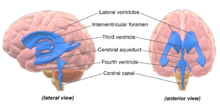
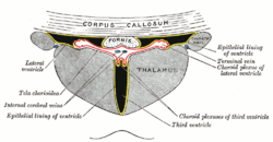
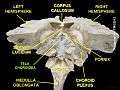
In the lateral ventricles the tela choroidea–a double-layered fold of pia mater and ependyma, produces the choroid fissure (also known as the choroidal fissure). The choroid fissure is C-shaped, runs between the fornix and the thalamus in the body of the ventricle, and between the stria terminalis and hippocampal fimbria in the inferior horn, and is the location of the attachment of the margins of the choroid plexus.[4][5] In the choroid fissure of the lateral ventricles, the tela choroidea is a lateral extension of the tela choroidea from the third ventricle.[6]
In the third ventricle the tela choroidea forms the roof of the ventricle. Two vascular fringes from the lower fold invaginate the roof and form the choroid plexus.[6]
The tela choroidea of the fourth ventricle (also known as the triangular lamella)[7] is a double layer of pia mater and ependyma, between the cerebellum and the lower part of the roof of the fourth ventricle. The two layers are continuous with each other in front, and are mostly adherent throughout. The anterior layer of the fold, contains vascular fringes which make up the choroid plexus.[6] The anterior layer is continuous inferiorly with the pia mater on the inferior cerebellar peduncles and the closed part of the medulla oblongata.
The posterior layer covers the antero-inferior surface of the cerebellum.
Blood supply
The blood supply of these plexuses is from the posterior inferior cerebellar artery. The lateral ventricles also contains the right and left internal cerebral veins (which drain the choroid plexuses) at its roof (the two veins unite to form the great cerebral vein).
The arteries carrying blood into the choroid plexuses are:
- the anterior choroidal artery (branch from the internal carotid).
- the posterior choroidal artery (branch from the posterior cerebral artery).
Medial posterior choroidal branches run forward beneath the splenium of the corpus callosum, and supply the tela chorioidea of the third ventricle and the choroid plexus.
Images
 Tela choroidea
References
- Larsen, William J. (2001). Human embryology (3rd ed.). Philadelphia, Pa.: Churchill Livingstone. p. 424. ISBN 978-0-443-06583-5.
- Alberts, Daniel (2012). Dorland's illustrated medical dictionary (32nd ed.). Philadelphia, PA: Saunders/Elsevier. p. 1878. ISBN 978-1-4160-6257-8.
- Sadler, T. (2010). Langman's medical embryology (11th ed.). Philadelphia: Lippincott William & Wilkins. p. 305. ISBN 978-0-7817-9069-7.
- Zemmoura, I. (2012). "The choroidal fissure: Anatomy and surgical implications". Advances and Technical Standards in Neurosurgery. Advances and Technical Standards in Neurosurgery. 38. Springer, Vienna. pp. 97–113. doi:10.1007/978-3-7091-0676-1_5. ISBN 978-3-7091-0675-4. PMID 22592413.
- "choroidal fissure - Definition". www.mondofacto.com.
- Alberts, Daniel (2012). Dorland's illustrated medical dictionary (32nd ed.). Philadelphia, PA: Saunders/Elsevier. p. 1878. ISBN 978-1-4160-6257-8.
- Alberts, Daniel (2012). Dorland's illustrated medical dictionary (32nd ed.). Philadelphia, PA: Saunders/Elsevier. p. 910. ISBN 978-1-4160-6257-8.