Falx cerebelli
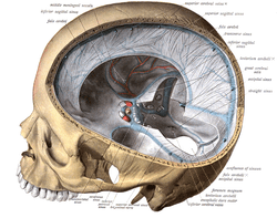
The falx cerebelli is a small sickle shaped fold of dura mater, projecting forwards into the posterior cerebellar notch as well as projecting into the vallecula of the cerebellum between the two cerebellar hemispheres.[1] The name comes from two Latin words: falx, meaning "curved blade or scythe", and cerebellum, meaning "brain".[2] Its base is attached, above, to the under and back part of the tentorium cerebelli; its posterior margin, to the lower division of the vertical crest on the inner surface of the occipital bone. The falx cerebelli generally lies somewhere between 2.8 and 4.5 cm in length and is approximately 1–2 mm thick.[3]
| Falx cerebelli | |
|---|---|
 Falx cerebelli seen in back portion of skull. | |
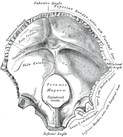 Occipital bone. Inner surface. (Portions "for faulx cerebelli" identified at center left.) | |
| Details | |
| Part of | Meninges |
| Identifiers | |
| Latin | Falx cerebelli |
| NeuroNames | 1238 |
| TA | A14.1.01.106 |
| FMA | 83974 |
| Anatomical terms of neuroanatomy | |
Variations in morphology
In its lower portion the falx cerebelli diminishes very rapidly in height and as it descends, it can divide into two smaller folds or diverging limbs,[4] which are lost on the sides of the foramen magnum. Other variations such as duplication,[5] triplication,[6] absence,[7] and fenestration are much less common. As dural venous sinuses are concurrent with the development of dural folds, duplication of the falx cerebelli is usually associated with duplicated occipital sinus.[8] Knowledge of these variations is important in preventing iatrogenic injuries in this region.
See also
- Falx (disambiguation) — other parts of the anatomy with names including "falx"
References
This article incorporates text in the public domain from page 874 of the 20th edition of Gray's Anatomy (1918) This article incorporates text from a public domain edition of Sobotta's Anatomy.
- http://babel.hathitrust.org/cgi/pt?q1=falx%20cerebelli;id=ien.35558004773517;seq=329;start=1;size=25;page=search;num=189
- Sihler, Andrew L. (1995). New Comparative Grammar of Greek and Latin. Oxford University Press. ISBN 978-0-19-508345-3. Retrieved 12 March 2013.
- Shoja MM, Tubbs RS, Khaki AA, Shokouhi G. A rare variation of the posterior cranial fossa: duplicated falx cerebelli, occipital venous sinus, and internal occipital crest. Folia Morphol (Warsz) 2006;65(2):171–173.
- http://babel.hathitrust.org/cgi/pt?q1=falx%20cerebelli;id=ien.35558004773517;seq=329;start=1;size=25;page=search;num=189
- Shoja MM, Tubbs RS, Khaki AA, Shokouhi G. A rare variation of the posterior cranial fossa: duplicated falx cerebelli, occipital venous sinus, and internal occipital crest. Folia Morphol (Warsz) 2006;65(2):171–173.
- Shoja MM, Tubbs RS, Loukas M, Shokouhi G, Oakes WJ. A complex dural-venous variation in the posterior cranial fossa: a triplicate falx cerebelli and an aberrant venous sinus. Folia Morphol (Warsz) 2007;66(2):148–51.
- Tubbs Rs, Dockery SE, Salter G, Elton S, Blount JP, Grabb PA, Oakes WJ. Absence of the falx cerebelli in a Chiari II malformation. Clin Anat. 2002;15(3):193–195.
- Shoja MM, Tubbs RS, Shokouhi GH, Ashrafian A, Oakes WJ. Abstract presented at the 23rd Annual Meeting of the American Association of Clinical Anatomists. Milwaukee, Wisconsin: 2006. A triple dural-venous variation in the posterior cranial fossa: A duplicated plus accessory falx cerebelli and an aberrant venous sinus.