Epidural space
Dura mater is the outermost meningeal layer that covers the brain and spinal cord. It consists of two layers; the inner meningeal layer and the outer periosteal layer. The potential space between the layers of the dura mater is called the epidural space. The epidural space exists around both the brain and the spinal cord.[1][2]
| Epidural space | |
|---|---|
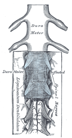 The medulla spinalis and its membranes | |
| Details | |
| Identifiers | |
| Latin | Spatium epidurale, spatium extradurale, cavum epidurale |
| MeSH | D004824 |
| TA | A14.1.01.112 |
| FMA | 71228 |
| Anatomical terminology | |
The anatomy term "epidural space" has its origin in the Ancient Greek language; ἐπί, "on, upon" + dura mater also known as "epidural cavity", "extradural space" or "peridural space". In humans the epidural space contains lymphatics, spinal nerve roots, loose connective tissue, adipose tissue, small arteries, dural venous sinuses and a network of internal vertebral venous plexuses.[3]
Cranial epidural space
In the skull, the periosteal layer of the dura mater adheres to the inner surface of the skull bones while the meningeal layer lays over the arachnoid mater. Between them is the cranial epidural space. The two layers of the dura mater separate at several places, with the meningeal layer projecting deeper into the brain parenchyma forming fibrous septa that compartmentalize the brain tissue. At these sites, the epidural space is wide enough to house the epidural venous sinuses.[2][4][5]
There are four fibrous septa:[4]
- Falx cerebri, that separates the left and right hemispheres of the cerebrum. It contains the superior sagittal sinus and inferior sagittal sinus.
- Tentorium cerebelli, which separates the cerebrum from cerebellum and contains the transverse sinus, straight sinus and superior petrosal sinus.
- Diaphragma sellae, that encloses the hypophyseal fossa from the superior side, cushioning the pituitary gland. It contains the anterior and posterior intercavernous sinuses.
- Falx cerebelli, which separates the left and right cerebellar hemispheres and contains the occipital sinus.
In pathological conditions fluid such as blood can fill this space. For example a torn meningeal artery (often the middle meningeal artery) or dural venous sinus (rarely) may bleed into this potential space and result in an epidural hematoma.[5]
Spinal epidural space
In the spinal canal, the periosteal layer adheres to the inner surface of the spinal canal which is formed by the bodies of vertebrae. The meningeal layer lays over the spinal arachnoid mater.[2] Between the two layers is the spinal epidural space. Unlike the cranial epidural space, the spinal epidural space contains adipose tissue and the external vertebral venous plexuses.[1]
References
- Waxman, Stephen G. (2010). Clinical neuroanatomy (26th ed.). New York: McGraw-Hill Medical. ISBN 9780071603997. OCLC 435703701.
- Blumenfeld, Hal (2010). Neuroanatomy through clinical cases (2nd ed.). Sunderland, Mass.: Sinauer Associates. ISBN 9780878930586. OCLC 473478856.
- Richardson, Jonathan; Groen, Gerbrand J. (2005-06-01). "Applied epidural anatomy". Continuing Education in Anaesthesia Critical Care & Pain. 5 (3): 98–100. doi:10.1093/bjaceaccp/mki026. ISSN 1743-1816.
- Patestas, Maria; Gartner, Leslie P. (2013). A Textbook of Neuroanatomy (1st ed.). New York, NY: John Wiley & Sons. ISBN 9781118687741. OCLC 899175403.
- Collins, Dawn; Goodfellow, John; Silva, Dulanka; Dardis, Ronan; Nagaraja, Sanjoy (2016). Neurology & neurosurgery. London: JP Medical Publishers. ISBN 9781907816741. OCLC 945569379.