Falx cerebri
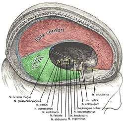
The falx cerebri is also known as the cerebral falx, named from its sickle-like form. It is a large, crescent-shaped fold of meningeal layer of dura mater that descends vertically in the longitudinal fissure between the cerebral hemispheres of the human brain. The falx cerebri attaches anteriorly at the crista galli in proximity to the cribriform plate and to the frontal and ethmoid sinuses. Posteriorly, it is connected with the upper surface of the cerebellar tentorium. Its superior margin is attached at midline to the internal surface of skull, as far back as the internal occipital protuberance. The superior sagittal sinus is contained in the superior margin of the falx cerebri and overlies the longitudinal fissure of the brain. The inferior sagittal sinus is contained in the inferior margin of the falx cerebri and arches over the corpus callosum, deep in the longitudinal fissure.[1]
| Falx cerebri | |
|---|---|
 | |
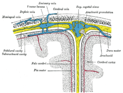 Diagrammatic representation of a section across the top of the skull, showing the membranes of the brain, etc. (Falx cerebri is yellow line running down center.) | |
| Details | |
| Part of | Meninges |
| Identifiers | |
| Latin | Falx cerebri |
| NeuroNames | 1237 |
| TA | A14.1.01.103 |
| FMA | 83967 |
| Anatomical terms of neuroanatomy | |
Calcification
Calcification of the falx cerebri is more prevalent in older patients, often without a determinable cause, and without pathogenic symptoms.[2]
Meningioma
Falcine meningioma is a meningioma arising from the falx cerebri and completely concealed by the overlying cortex. Falcine meningioma tends to grow predominately into one cerebral hemisphere but is often bilateral, and in some patients the tumor grows into the inferior edge of the sagittal sinus. However, although much information is available regarding meningiomas, little is known about falcine meningiomas.[3]
Additional images
 Falx cerebri in relation to the skull.
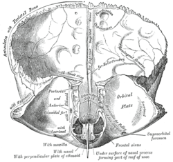 Frontal bone. Inner surface.
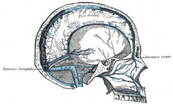 Sagittal section of the skull, showing the sinuses of the dura.
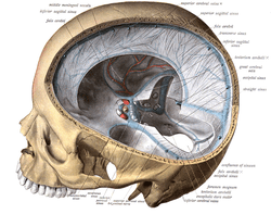
_description.JPG) Human brain dura mater (reflections)
Human brain dura mater (reflections) Falx cerebri
See also
- Falx (disambiguation)—other parts of the anatomy with names including "falx"
References
This article incorporates text in the public domain from page 873 of the 20th edition of Gray's Anatomy (1918)
- Saladin K. "Anatomy & Physiology: The Unity of Form and Function. New York: McGraw Hill, 2014. Print. pp 512, 770-773
- Daghighi MH, Rezaei V, Zarrintan S, Pourfathi H (2007). "Intracranial physiological calcifications in adults on computed tomography in Tabriz, Iran." Folia Morphol (Warsz). 66 (2):115-9. PMID 17594669
- Chung SB, Kim CY, Park CK, Kim DG, Jung HW (2007). "Falx Meningiomas: Surgical Results and Lessons Learned from 68 Cases." J Korean Neurosurg Soc. 42 (4): 276-280. PMC 2588203
External links
| Wikimedia Commons has media related to Falx cerebri. |
- Anatomy photo:28:st-1602 at the SUNY Downstate Medical Center
- MedEd at Loyola grossanatomy/dissector/labs/h_n/cranium/cn1_1a.htm
| Authority control |
|---|