Pterygopalatine fossa
In human anatomy, the pterygopalatine fossa (sphenopalatine fossa) is a fossa in the skull. A human skull contains two pterygopalatine fossae—one on the left side, and another on the right side. Each fossa is a cone-shaped paired depression deep to the infratemporal fossa and posterior to the maxilla on each side of the skull, located between the pterygoid process and the maxillary tuberosity close to the apex of the orbit.[1] It is the indented area medial to the pterygomaxillary fissure leading into the sphenopalatine foramen. It communicates with the nasal and oral cavities, infratemporal fossa, orbit, pharynx, and middle cranial fossa through eight foramina.[2]
| Pterygopalatine fossa | |
|---|---|
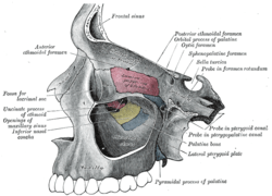 Left maxillary sinus opened from the exterior. | |
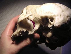 Human skull with entrance to pterygopalatine fossa marked in purple | |
| Details | |
| Identifiers | |
| Latin | fossa pterygopalatina |
| MeSH | D056739 |
| TA | A02.1.00.025 |
| FMA | 75309 |
| Anatomical terms of bone | |
Structure
Boundaries
It has the following boundaries:
- anterior: superomedial part of the infratemporal surface of maxilla
- posterior: root of the pterygoid process and adjoining anterior surface of the greater wing of sphenoid bone
- medial: perpendicular plate of the palatine bone and its orbital and sphenoidal processes
- lateral: pterygomaxillary fissure
- inferior: part of the floor is formed by the pyramidal process of the palatine bone.
Passages
The following passages connect the fossa with other parts of the skull:
| Direction | Passage | Connection |
|---|---|---|
| Posteriorly | foramen rotundum | middle cranial fossa |
| pterygoid canal (Vidian) | middle cranial fossa, foramen lacerum | |
| palatovaginal canal (pharyngeal) | nasal cavity/nasopharynx | |
| Anteriorly | inferior orbital fissure | orbit |
| Medially | sphenopalatine foramen | nasal cavity |
| Laterally | pterygomaxillary fissure | infratemporal fossa |
| Inferiorly | greater palatine canal (pterygopalatine) | oral cavity, |
Functions
The pterygopalatine fossa contains
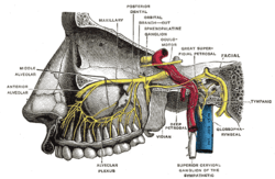
- the pterygopalatine ganglion suspended by nerve roots from the maxillary nerve
- the terminal third of the maxillary artery
- the maxillary nerve (CN V2, the second division of the trigeminal nerve), with which is the nerve of the pterygoid canal, a combination of the greater petrosal nerve (preganglionic parasympathetic) and the deep petrosal nerve (postganglionic sympathetic). To obtain block anesthesia of the entire second division of the trigeminal nerve, an intraoral injection can be administered into this area.
See also
- Pterygopalatine canal (disambiguation)
- Fossa in the Human Body
Additional images
 Alveolar branches of superior maxillary nerve and pterygopalatine ganglion
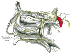 The pterygopalatine ganglion and its branches
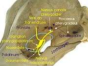 Pterygopalatine fossa in a dog
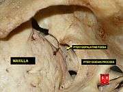 Pterygopalatine fossa
References
- Illustrated Anatomy of the Head and Neck, Fehrenbach and Herring, Elsevier, 2012, page 69
- Osborn, Anne (March 1979). "Radiology of the Pterygoid Plates and Pterygopalatine Fossa" (PDF). AJR. 132 (3): 389–394. doi:10.2214/ajr.132.3.389. PMID 106641.