Mylohyoid muscle
The mylohyoid muscle is a paired muscle running from the mandible to the hyoid bone, forming the floor of the oral cavity of the mouth.[1] It is named after its two attachments near the molar teeth ("mylo" comes from the Greek word for "molar").[2] These muscles are mesodermal in embryologic origin. The mylohyoid muscle is derived from the first pharyngeal arch.
| Mylohyoid muscle | |
|---|---|
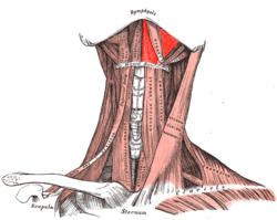 Muscles of the neck seen from the front (mylohyoid muscle colored in bright red) | |
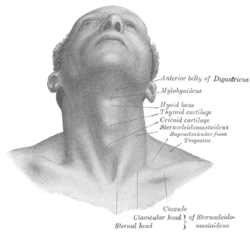 Surface anatomy of the neck seen from the front (mylohyoid muscle labeled at right, second from top) | |
| Details | |
| Origin | Mylohyoid line (mandible) |
| Insertion | Body of hyoid bone and median ridge |
| Artery | Mylohyoid branch of inferior alveolar artery and submental artery of facial artery |
| Nerve | Mylohyoid nerve, from inferior alveolar branch of mandibular nerve |
| Actions | Raises oral cavity floor, elevates hyoid, elevates tongue, depresses mandible |
| Identifiers | |
| Latin | Musculus mylohyoideus |
| TA | A04.2.03.006 |
| FMA | 46320 |
| Anatomical terms of muscle | |
Structure
The mylohyoid muscle is flat and triangular, and is situated immediately superior to the anterior belly of the digastric muscle. It is a pharyngeal muscle (derived from the first pharyngeal arch) and classified as one of the suprahyoid muscles. Together, the paired mylohyoid muscles form a muscular floor for the oral cavity of the mouth.[3]
The two mylohyoid muscles arise from the mandible at the mylohyoid line, which extends from the mandibular symphysis in front to the last molar tooth behind. The posterior fibers pass inferomedially and insert at anterior surface of the hyoid bone. The medial fibres of the two mylohyoid muscles unite in a midline raphe (where the two muscles intermesh).[4]
The mylohyoid muscle separates the sublingual space from the submandibular space, which communicate via a lateral gap between the mylohyoid and hyoglossus muscles at the posterior free margin of mylohyoid muscle.[5] The submandibular gland wraps around the edges of the mylohyoid, and is divided into superficial and deep lobes above and below the muscle.[6]
Innervation
The mylohyoid muscle is innervated by a branch of the mandibular nerve, the inferior alveolar nerve. The mylohyoid nerve is a branch of the inferior alveolar nerve. The mylohyoid nerve emerges to give motor supply to the mylohyoid muscle.[1]
Variations
The mylohyoid may be united to or replaced by the anterior belly of the digastric muscle; accessory slips to other hyoid muscles are frequent. This median raphé is sometimes absent; the fibers of the two muscles are then continuous.
An area of herniation of the sublingual gland, blood vessels, or fat, may be present, with studies reporting this in 10-50% of people.[5]
Function
The mylohyoid elevates the hyoid and the tongue. This is particularly important during swallowing and speaking. Alternatively, if other muscles are used to keep the position of the hyoid fixed, then the mylohyoid depresses the mandible.[1] It also functions as reinforcing the floor of mouth.[1]
Clinical relevance
The mylohyoid may be imaged by CT or MRI.[5] The mylohyoid separates the submandibular space below from the sublingual space above. Around the posterior border of mylohoid, these spaces communicate. Infections, especially odontogenic infections can spread from one space to the other via this communication, or alternatively penetrate the mylohyoid which is a poor barrier to the spread of infection. Because the attachment of mylohyoid (the mylohoid line) becomes more superior towards the posterior of the mandible, posterior infected teeth are more likely to drain into the mandibular space, and infected anterior teeth are more likely to drain into the sublingual space, since the apices of the teeth are more likely to be below and above the mylohoid line respectively (see diagram).
Additional images
 Anterior view
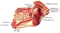 Medial view
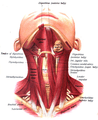
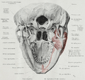 The origin of the mylohyoid muscle, inferior view.
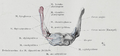 The insertion of the mylohyoid muscle on the hyoid bone.
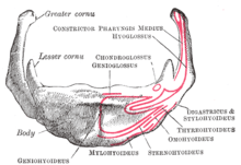 Hyoid bone. Anterior surface. Enlarged.
 Mylohyoid muscle
Notes
- Drake, Vogl & Tibbitts 2005, p. 987.
- http://www.anatomy.usyd.edu.au/glossary/glossary.cgi?page=m%5B%5D
- Herring & Fehrenbach 2013, p. 212.
- Drake, Vogl & Tibbitts 2005, pp. 987–8.
- Otonari-Yamamoto, Mika; Nakajima, Koh; Tsuji, Yuriko; Otonari, Takamichi; Curtin, Hugh D.; Okano, Tomohiro; Sano, Tsukasa (2010). "Imaging of the Mylohyoid Muscle: Separation of Submandibular and Sublingual Spaces". American Journal of Roentgenology. 194 (5): W431–8. doi:10.2214/AJR.09.3516. PMID 20410390.
- Drake, Vogl & Tibbitts 2005, p. 997.
References
- Drake, Richard L.; Vogl, Wayne; Tibbitts, Adam W.M. Mitchell (2005). Gray's anatomy for students. Philadelphia: Elsevier/Churchill Livingstone. ISBN 978-0-443-06612-2.
- Herring, Margaret J.; Fehrenbach, Susan W. (2013). Illustrated anatomy of the head and neck (4th ed.). St. Louis, MO: Elsevier/Saunders. ISBN 978-1-4377-2419-6.
- This article incorporates text in the public domain from page 393 of the 20th edition of Gray's Anatomy (1918)
External links
- "Anatomy diagram: 25420.000-1". Roche Lexicon - illustrated navigator. Elsevier. Archived from the original on 2014-01-01.
| Authority control |
|---|