Submandibular space
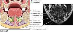
The submandibular space is a fascial space of the head and neck (sometimes also termed fascial spaces or tissue spaces). It is a potential space, and is paired on either side, located on the superficial surface of the mylohyoid muscle between the anterior and posterior bellies of the digastric muscle.[1] The space corresponds to the anatomic region termed the submandibular triangle, part of the anterior triangle of the neck.
| Submandibular space | |
|---|---|
.png) Right submandibular space situated on the superficial surface of mylohyoid muscle, between the anterior and posterior bellies of the digastric muscle (highlighted in green). | |
 | |
| Details | |
| Identifiers | |
| Latin | Spatium submandibulare |
| Anatomical terminology | |
Location and structure
Anatomic boundaries
The anatomic boundaries of each submandibular space are:[2]
- the mylohyoid muscle superiorly,
- the skin, superficial fascia, platysma muscle and superficial layer of the deep cervical fascia inferiorly and laterally,
- the medial surface of the mandible anteriorly and laterally,
- the hyoid bone posteriorly,
- the anterior belly of the digastric muscle medially.
Communications
The communications of the submandibular space are:
- medially and anteriorly to the submental space (located medial to the paired submandibular spaces, separated from them by the anterior bellies of the digastric muscles).
- posteriorly and superiorly to the sublingual space (located above the mylohyoid muscle)
- inferiorly to the lateral pharyngeal space
Contains
In health, the contents of the space are:
- the submandibular gland, which largely fills the space,
- branches of the facial and lingual artery
- lymph nodes.
- cranial nerve XII
- nerve to mylohyoid muscle
Clinical relevance
.png)
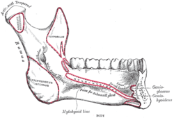
Infections may spread into the submandibular space, e.g. odontogenic infections, often related to the mandibular molar teeth. This is due to the fact that the attachment of mylohyoid (the mylohoid line) becomes more superior towards the posterior of the mandible, meaning that the roots of the posterior teeth are more likely to be below mylohyoid than above.
Signs and symptoms of a submandibular space infection might include trismus (difficulty opening the mouth), inability to palpate (feel) the inferior border of the mandible and swelling of the face over the submandibular region.
If the space contains pus, the usual treatment is by incision and drainage. The site of the incision is extra-oral, and usually made 2–3 cm below, and parallel to, the inferior border of the mandible.
Ludwig's angina is a serious infection involving the submandibular, sublingual and submental spaces bilaterally.[3] Ludwig's angina may extend into the pharyngeal and cervical spaces, and the swelling can compress the airway and cause dyspnoea (difficulty breathing).[3]
References
- Contributing illustrators Craig JA [et al.], Norton N; illustrated by Netter FH; (2007). Netter's head and neck anatomy for dentistry. Philadelphia, Pa.: Saunders Elsevier. p. 466. ISBN 9781929007882.CS1 maint: extra punctuation (link)
- Mani, K George Varghese, foreword by Varghese (2008). A practical guide to hospital dentistry. New Delhi: Jaypee Bros. Medical Pub. p. 62. ISBN 9788184482430.
- Kenneth M. Hargreaves, Stephen Cohen; web editor, Louis H.Berman, eds. (2010). Cohen's pathways of the pulp (10th ed.). St. Louis, Mo.: Mosby Elsevier. pp. 590, 592. ISBN 978-0323064897.CS1 maint: uses editors parameter (link)