Hyoglossus
The hyoglossus, thin and quadrilateral, arises from the side of the body and from the whole length of the greater cornu of the hyoid bone, and passes almost vertically upward to enter the side of the tongue, between the styloglossus and the inferior longitudinal muscle of the tongue. It forms a part of the floor of submandibular triangle.
| Hyoglossus | |
|---|---|
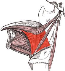 Extrinsic muscles of the tongue. Left side. (Hyoglossus visible at center.) | |
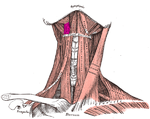 Muscles of the neck. Anterior view. Hyoglossal muscle in purple | |
| Details | |
| Origin | Hyoid |
| Insertion | side of the tongue |
| Nerve | Hypoglossal (CN XII) |
| Actions | depresses and retracts the tongue |
| Identifiers | |
| Latin | musculus hyoglossus |
| TA | A05.1.04.102 |
| FMA | 46691 |
| Anatomical terms of muscle | |
Structure
The fibers arising from the body of the hyoid bone overlap those from the greater cornu.
Structures that are medial/deep to the hyoglossus are the glossopharyngeal nerve (cranial nerve 9), the stylohyoid ligament and the lingual artery and lingual vein.
The lingual vein passes medial to the hyoglossus, and the lingual artery passes deep to the hyoglossus. Laterally, in between the hyoglossus muscle and the mylohyoid muscle lay several important structures (from upper to lower): sublingual gland, submandibular duct, lingual nerve, vena comitans of hypoglossal nerve, and the hypoglossal nerve. Note, posteriorly, the lingual nerve is superior to the submandibular duct and a portion of the submandibular salivary gland protrudes into the space between the hyoglossus and mylohyoid muscles.
Function
The hyoglossus depresses and retracts the tongue and makes the dorsum more convex.
Additional images
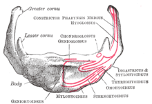 Hyoid bone. Anterior surface. Enlarged.
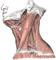 Muscles of the neck. Lateral view.
 The internal carotid and vertebral arteries. Right side.
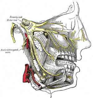 Distribution of the maxillary and mandibular nerves, and the submaxillary ganglion.
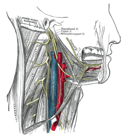 Hypoglossal nerve, cervical plexus, and their branches.
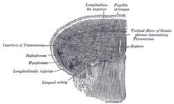 Coronal section of tongue, showing intrinsic muscles.
 Hyoglossus Muscle
References
This article incorporates text in the public domain from page 1129 of the 20th edition of Gray's Anatomy (1918)
External links
- Anatomy figure: 34:02-09 at Human Anatomy Online, SUNY Downstate Medical Center
- "Anatomy diagram: 25420.000-1". Roche Lexicon - illustrated navigator. Elsevier. Archived from the original on 2014-01-01.
- Diagram
| Authority control |
|---|