Lateral pterygoid muscle
The lateral pterygoid or external pterygoid is a muscle of mastication with two heads. It lies superiorly to the medial pterygoid.
| Lateral pterygoid muscle | |
|---|---|
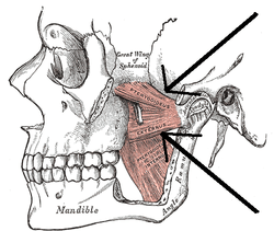 The Pterygoidei; the zygomatic arch and a portion of the ramus of the mandible have been removed (labeled as "pterygoideus externus", visible in pink at center) | |
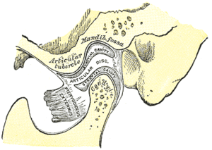 Sagittal section of the temporomandibular joint (labeled as "pterygoideus externus", visible in gray at bottom right) | |
| Details | |
| Origin | Superior head: infratemporal fossa. Inferior head: lateral pterygoid plate |
| Insertion | Superior head: anterior side of the condyle. Inferior head: pterygoid fovea |
| Artery | Pterygoid branches of maxillary artery |
| Nerve | Lateral pterygoid nerve from the mandibular nerve |
| Actions | depresses mandible, protrude mandible, side to side movement of mandible |
| Identifiers | |
| Latin | Musculus pterygoideus lateralis, musculus pterygoideus externus |
| TA | A04.1.04.006 |
| FMA | 49015 |
| Anatomical terms of muscle | |
Origin and insertion
The upper/superior head originates on the infratemporal surface and infratemporal crest of the greater wing of the sphenoid bone and inserts onto the articular disc and fibrous capsule of the temporomandibular joint.
The lower/inferior head originates on the lateral surface of the lateral pterygoid plate and inserts onto the neck of condyloid process of the mandible; upper/superior head
Innervation
The mandibular branch of the fifth cranial nerve, the trigeminal nerve, specifically the lateral pterygoid nerve, innervates the lateral pterygoid muscle.
Function
The primary function of the lateral pterygoid muscle is to pull the head of the condyle out of the mandibular fossa along the articular eminence to protrude the mandible. A concerted effort of the lateral pterygoid muscles helps in lowering the mandible and opening the jaw, whereas unilateral action of a lateral pterygoid produces contralateral excursion (a form of mastication), usually performed in concert with the medial pterygoids.
Unlike the other three muscles of mastication, the lateral pterygoid is the only muscle of mastication that assists in depressing the mandible (opening the jaw). At the beginning of this action it is assisted by the digastric, mylohyoid and geniohyoid muscles.
Additional images
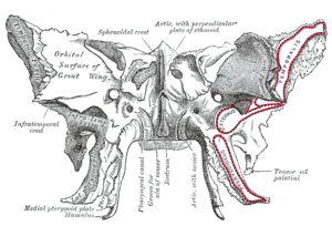 Sphenoid bone. Anterior and inferior surfaces.
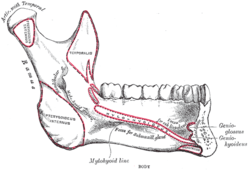 Mandible. Inner surface. Side view.
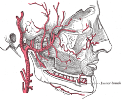 Plan of branches of internal maxillary artery.
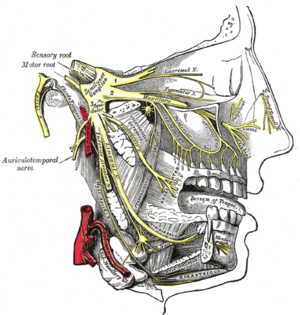 Distribution of the maxillary and mandibular nerves, and the submaxillary ganglion.
External links
- lesson4 at The Anatomy Lesson by Wesley Norman (Georgetown University) (musclesofmastication2)
- MedicalMnemonics.com: 70
- "Anatomy diagram: 25420.000-1". Roche Lexicon - illustrated navigator. Elsevier. Archived from the original on 2014-01-01.
- Cross section at tufts.edu
| Authority control |
|---|