Palatopharyngeus muscle
The palatopharyngeus (palatopharyngeal or pharyngopalatinus) muscle is a small muscle in the roof of the mouth.
| Palatopharyngeus muscle | |
|---|---|
 | |
| Details | |
| Origin | Palatine aponeurosis and hard palate |
| Insertion | Upper border of thyroid cartilage (blends with constrictor fibers) |
| Artery | Facial artery |
| Nerve | Pharyngeal branch of vagus nerve |
| Actions | Pulls pharynx and larynx upward |
| Identifiers | |
| Latin | Musculus palatopharyngeus |
| TA | A05.2.01.105 |
| FMA | 46666 |
| Anatomical terms of muscle | |
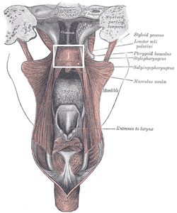
It is a long, fleshy fasciculus, narrower in the middle than at either end, forming, with the mucous membrane covering its surface, the palatopharyngeal arch.
Structure
It is separated from the palatoglossus muscle by an angular interval, in which the palatine tonsil is lodged. It arises from the soft palate, where it is divided into two fasciculi by the levator veli palatini and musculus uvulae.
- The posterior fasciculus lies in contact with the mucous membrane, and joins with that of the opposite muscle in the middle line.
- The anterior fasciculus, the thicker, lies in the soft palate between the levator and tensor veli palatini muscles, and joins in the middle line the corresponding part of the opposite muscle.
Passing laterally and downward behind the palatine tonsil, the palatopharyngeus joins the stylopharyngeus and is inserted with that muscle into the posterior border of the thyroid cartilage, some of its fibers being lost on the side of the pharynx and others passing across the middle line posteriorly to decussate with the muscle of the opposite side.
Innervation
Motor innervation of this muscle is provided through the pharyngeal plexus of the CN X (vagal nerve), SVE (special visceral efferent) fibers.
Function
The palatine velum is slightly raised by the levator veli palatini and made tense by the tensor veli palatini; the palatopharyngeus muscles, by their contraction, pull the pharynx upward over the bolus of food and nearly come together, the uvula filling up the slight interval between them.
By these means the bolus is prevented from passing into the nasopharynx; at the same time, the palatopharyngeus muscles form an inclined plane, directed obliquely downward and backward, along the under surface of which the bolus descends into the lower part of the pharynx.
Additional images
 Cadaver dissection showing the palatopharyngeus muscle
References
This article incorporates text in the public domain from page 1140 of the 20th edition of Gray's Anatomy (1918)
External links
| Wikimedia Commons has media related to Palatopharyngeus muscle. |
- Anatomy figure: 31:04-05 at Human Anatomy Online, SUNY Downstate Medical Center
- Anatomy photo:34:04-0104 at the SUNY Downstate Medical Center
- "Anatomy diagram: 05287.011-1". Roche Lexicon - illustrated navigator. Elsevier. Archived from the original on 2014-01-01.
| Authority control |
|---|