Caliciviridae
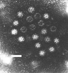
The Caliciviridae are a family of viruses, members of Class IV of the Baltimore scheme. They are positive-sense, single-stranded RNA which is not segmented[1]. Thirteen species are placed in this family, divided among eleven genera.[2] Diseases associated with this family include feline calicivirus (respiratory disease), rabbit hemorrhagic disease virus (often-fatal hemorrhages), and Norwalk group of viruses (gastroenteritis).[2][3] Caliciviruses naturally infect vertebrates, and have been found in a number of organisms such as humans, cattle, pigs, cats, chickens, reptiles, dolphins and amphibians. The caliciviruses have a simple construction and are not enveloped. The capsid appears hexagonal/spherical and has icosahedral symmetry (T=1 or T=3[3]) with a diameter of 35–39 nm.[4]
| Caliciviridae | |
|---|---|
 | |
| Virus classification | |
| (unranked): | Virus |
| Realm: | Riboviria |
| Phylum: | incertae sedis |
| Family: | Caliciviridae |
| Genera | |
Caliciviruses are not very well studied because until recently, they could not be grown in culture, and no suitable animal model has been found. However, the recent application of modern genomic technologies has led to an increased understanding of the virus family.[4] A recent isolate from rhesus monkeys—Tulane virus—can be grown in culture, and this system promises to increase understanding of these viruses.[5]
The name calicivirus is derived from the Greek word calyx meaning cup or goblet, due to many strains having visible cup-shaped depressions.
Taxonomy
The Caliciviridae family includes these genera:
- Genus Lagovirus; type species: Rabbit hemorrhagic disease virus
- Genus Nebovirus; type species: Newbury-1 virus
- Genus Norovirus; type species: Norwalk virus
- Genus Sapovirus; type species: Sapporo virus
- Genus Vesivirus; type species: Vesicular exanthema of swine virus
Group: ssRNA(+)
Two additional genera have been proposed: Recovirus for a novel calicivirus detected in stool specimens (Tulane virus) from rhesus monkeys[6] and Valovirus for a novel group of swine caliciviruses known as the St-Valérien-like viruses. These genera have yet to be officially approved. A number of other caliciviruses remain unclassified, including the chicken calicivirus.
Virology
All viruses in this family possess a nonsegmented, polyadenylated, positive-sense, single-strand RNA genome around 7.5–8.5 kilobases in length, enclosed in an icosahedral capsid of 27–40 nanometers in diameter.
| Genus | Structure | Symmetry | Capsid | Genomic arrangement | Genomic segmentation |
|---|---|---|---|---|---|
| Nebovirus | Icosahedral | T=1, T=3 | Non-enveloped | Linear | Monopartite |
| Lagovirus | Icosahedral | T=1, T=3 | Non-enveloped | Linear | Monopartite |
| Sapovirus | Icosahedral | T=3 | Non-enveloped | Linear | Monopartite |
| Norovirus | Icosahedral | T=1, T=3 | Non-enveloped | Linear | Monopartite |
| Vesivirus | Icosahedral | T=1, T=3 | Non-enveloped | Linear | Monopartite |
Life cycle
Viral replication is cytoplasmic. Entry into the host cell is achieved by attachment to host receptors, which mediate endocytosis. Replication follows the positive-stranded RNA virus replication model. Positive-stranded RNA virus transcription is the method of transcription. Translation takes place by leaky scanning, and RNA termination-reinitiation. Vertebrates serve as the natural host. Transmission routes are fecal-oral.[3]
| Genus | Host details | Tissue tropism | Entry details | Release details | Replication site | Assembly site | Transmission |
|---|---|---|---|---|---|---|---|
| Nebovirus | Bovine | Liver | Cell receptor endocytosis | Lysis | Cytoplasm | Cytoplasm | Unknown |
| Lagovirus | Lagomorphs | Liver | Cell receptor endocytosis | Lysis | Cytoplasm | Cytoplasm | Contact |
| Sapovirus | Humans; swine | Intestinal epithelium | Cell receptor endocytosis | Lysis | Cytoplasm | Cytoplasm | Oral-fecal |
| Norovirus | Humans; mammals | Intestinal epithelium | Cell receptor endocytosis | Lysis | Cytoplasm | Cytoplasm | Oral-fecal |
| Vesivirus | Felines | Upper respiratory | Cell receptor endocytosis | Lysis | Cytoplasm | Cytoplasm | Aerosol |
Human disease
Calicivirus infections commonly cause acute gastroenteritis, which is the inflammation of the stomach and intestines (e.g. the Norwalk virus). Symptoms can include vomiting and diarrhea. These symptoms emerge after an incubation time of 2 days and the symptoms only generally last for 3 days. Most calicivirus infections do not call for medical attention, but those who are immunocompromised may need to be hospitalized for rehydration therapy.
Animal viruses
Feline calicivirus (FCV)—a member of the Vesivirus—represents an important pathogen of cats.
Sapovirus, Norovirus, and Vesivirus have been detected in pigs, making this animal species of particular interest in the study of calicivirus pathogenesis and host range.
The first mouse norovirus, murine norovirus 1 (MNV-1), was discovered in 2003. Since then, numerous murine norovirus strains have been identified and they were assigned a new genogroup in the genus Norovirus.
Rabbit hemorrhagic disease virus is a pathogen of rabbits that causes major problems throughout the world where rabbits are reared for food and clothing, make a significant contribution to ecosystem ecology, and where they support valued wildlife as a food source.[4]
Note
Australia and New Zealand, in an effort to control their rabbit populations, have intentionally spread calicivirus.
References
- Vinjé, J; Estes, MK; Esteves, P; Green, KY; Katayama, K; Knowles, NJ; L'Homme, Y; Martella, V; Vennema, H; White, PA; ICTV Report Consortium (November 2019). "ICTV Virus Taxonomy Profile: Caliciviridae". The Journal of General Virology. 100 (11): 1469–1470. doi:10.1099/jgv.0.001332. PMID 31573467.
- "ICTV Report Caliciviridae".
- "Viral Zone". ExPASy. Retrieved 15 June 2015.
- Hansman, GS (editor) (2010). Caliciviruses: Molecular and Cellular Virology. Caister Academic Press. ISBN 978-1-904455-63-9.CS1 maint: extra text: authors list (link)
- Yu G, Zhang D, Guo F, Tan M, Jiang X, Jiang W (2013) Cryo-EM structure of a novel calicivirus, Tulane Virus. PLoS One 8(3):e59817. doi: 10.1371/journal.pone.0059817
- Farkas T, Sestak K, Wei C, Jiang X (2008) Characterization of a rhesus monkey calicivirus representing a new genus of Caliciviridae. J Virol 82: 5408–5416. doi: 10.1128/JVI.00070-08
External links
- ICTV Report: Caliciviridae
- Caliciviridae description page from the International Committee on Taxonomy of Viruses site
- MicrobiologyBytes: Caliciviruses
- Human caliciviruses
- Stanford University
- Virus Pathogen Database and Analysis Resource (ViPR): Caliciviridae
- Viralzone: Caliciviridae
- 3D macromolecular structures of Caliciviridae from the EM Data Bank(EMDB)
- ICTV