Bartholin's gland
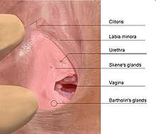
The Bartholin's glands (also called Bartholin glands or greater vestibular glands) are two pea sized compound alveolar glands[2] located slightly posterior and to the left and right of the opening of the vagina. They secrete mucus to lubricate the vagina and are homologous to bulbourethral glands in males. However, while Bartholin's glands are located in the superficial perineal pouch in females, bulbourethral glands are located in the deep perineal pouch in males. Their duct length is 1.5 to 2.0 cm and they open into navicular fossa.[2] The ducts are paired and they open on the surface of the vulva.
| Bartholin's gland | |
|---|---|
 Female genital organs | |
| Details | |
| Precursor | Urogenital sinus |
| Artery | external pudendal artery[1] |
| Nerve | ilioinguinal nerve[1] |
| Lymph | superficial inguinal lymph nodes |
| Identifiers | |
| Latin | glandula vestibularis major |
| MeSH | D001472 |
| TA | A09.2.01.016 |
| FMA | 9598 |
| Anatomical terminology | |
History

Bartholin's glands were first described in the 17th century by the Danish anatomist Caspar Bartholin the Younger (1655–1738).[3][4] Some sources mistakenly ascribe their discovery to his grandfather, theologian and anatomist Caspar Bartholin the Elder (1585–1629).[5]
Function
Bartholin's glands secrete mucus to provide vaginal lubrication during sexual arousal.[4][6][7] The fluid may slightly moisten the labial opening of the vagina, serving to make contact with this sensitive area more comfortable.[8]
Clinical pathology
It is possible for the Bartholin's glands to become blocked and inflamed resulting in pain.[8] This is known as bartholinitis or a Bartholin's cyst.[4][9] A Bartholin's cyst in turn can become infected and form an abscess. Adenocarcinoma of the gland is rare and benign tumors and hyperplasia are even more rare.[10] Bartholin gland carcinoma is a rare malignancy that occurs in 1% of vulvar cancers. This may be due to the presence of three different types of epithelial tissue.[3] Inflammation of the Skene's glands and Bartholin glands may appear similar to cystocele.[11]
See also
- Skene's gland
- Thomas Bartholin
References
- Greater Vestibular (Bartholin) gland Archived January 12, 2007, at the Wayback Machine
- Manual of Obstetrics. (3rd ed.). Elsevier. pp. 1-16. ISBN 9788131225561.
- Heller, Debra S.; Bean, Sarah (2014). "Lesions of the Bartholin Gland". Journal of Lower Genital Tract Disease. 18 (4): 351–357. doi:10.1097/LGT.0000000000000016. ISSN 1089-2591.
- Lee, M. Y; Dalpiaz, A; Schwamb, R; Miao, Y; Waltzer, W; Khan, A (2015). "Clinical Pathology of Bartholin's Glands: A Review of the Literature". Current Urology. 8 (1): 22–25. doi:10.1159/000365683. PMC 4483306.
- C. C. Gillispie (ed.): Dictionary of Scientific Biography, New York 1970..
- "Viscera of the Urogenital Triangle". University of Arkansas Medical School.
- Chrétien, F.C.; Berthou J. (September 18, 2006). "Crystallographic investigation of the dried exudate of the major vestibular (Bartholin's) glands in women". Eur J Obstet Gynecol Reprod Biol. 135 (1): 116–22. doi:10.1016/j.ejogrb.2006.06.031. PMID 16987591.
- "Bartholin's Gland". Discovery Health. Archived from the original on 2008-08-04.
- Sue E. Huether (2014). Pathophysiology: The Biologic Basis for Disease in Adults and Children. Elsevier Health Sciences. p. 817. ISBN 9780323293754.
- Argenta PA, Bell K, Reynolds C, Weinstein R (Oct 1997). "Bartholin's gland hyperplasia in a postmenopausal woman". Obstetrics & Gynecology. 90 (4 part 2): 695–7. doi:10.1016/S0029-7844(97)00409-2. PMID 11770602.
- "Cystoceles, Urethroceles, Enteroceles, and Rectoceles - Gynecology and Obstetrics - Merck Manuals Professional Edition". Merck Manuals Professional Edition. Retrieved 2018-02-06.
| Authority control |
|---|