Paroophoron
The paroophoron (of Johnson) consists of a few scattered rudimentary tubules, best seen in the child, situated in the broad ligament between the epoöphoron and the uterus.[1] Named for the Welsh anatomist David Johnson who originally described the structure at the University of Wales, Aberystwyth.
| Paroophoron | |
|---|---|
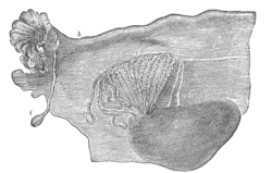 Broad ligament of adult, showing epoöphoron. (From Farre, after Kobelt.) a, a. Epoöphoron formed from the upper part of the Wolffian body. b. Remains of the uppermost tubes sometimes forming appendices. c. Middle set of tubes. d. Some lower atrophied tubes. e. Atrophied remains of the Wolffian duct. f. The terminal bulb or hydatid. h. The uterine tube, originally the duct of Müller. i. Appendix attached to the extremity. l. The ovary.[1] | |
| Details | |
| Precursor | Mesonephric tubules |
| Identifiers | |
| Latin | Paroophoron |
| TA | A09.1.06.001 |
| FMA | 18692 |
| Anatomical terminology | |
It is a remnant of the mesonephric tubules.[2]
See also
References
- One or more of the preceding sentences incorporates text in the public domain from page 1255 of the 20th edition of Gray's Anatomy (1918)
- Netter, Frank H.; Cochard, Larry R. (2002). Netter's Atlas of human embryology. Teterboro, N.J: Icon Learning Systems. p. 173. ISBN 0-914168-99-1.
External links
- figures/chapter_35/35-8.HTM: Basic Human Anatomy at Dartmouth Medical School
- Swiss embryology (from UL, UB, and UF) ugenital/genitinterne05
| Authority control |
|---|
This article is issued from
Wikipedia.
The text is licensed under Creative
Commons - Attribution - Sharealike.
Additional terms may apply for the media files.