Epoophoron
The epoophoron or epoöphoron (also called organ of Rosenmüller[2][3] or the parovarium) is a remnant of the mesonephric tubules that can be found next to the ovary and fallopian tube.
| Epoophoron | |
|---|---|
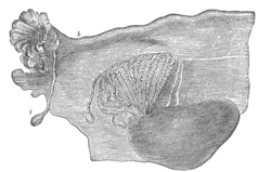 Broad ligament of adult, showing epoöphoron. (From Farre, after Kobelt.) a, a. Epoöphoron formed from the upper part of the Mesonephric body. b. Remains of the uppermost tubes sometimes forming appendices. c. Middle set of tubes. d. Some lower atrophied tubes. e. Atrophied remains of the Mesonephric duct. f. The terminal bulb or hydatid. h. The uterine tube, originally the duct of Müller. i. Appendix attached to the extremity. l. The ovary. | |
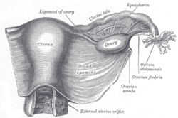 Uterus and right broad ligament, seen from behind. The epoophoron is visible in upper right | |
| Details | |
| Precursor | mesonephric duct[1] |
| Identifiers | |
| TA | A09.1.05.001 |
| FMA | 18691 |
| Anatomical terminology | |
Anatomy
It may contain 10–15 transverse small ducts or tubules that lead to the Gartner’s duct (also longitudinal duct of epoophoron) that represents the caudal remnant of the mesonephric duct and passes through the broad ligament and the lateral wall of the cervix and vagina.
The epoophoron is a homologue to the epididymis in the male.
While the epoophoron is located in the lateral portion of the mesosalpinx and mesovarium, the paroophoron (residual remnant of that part of the mesonephric duct that forms the paradidymis in the male) lies more medially in the mesosalpinx.
Clinical significance
Clinically the organ may give rise to a local paraovarian cyst or adenoma.
References
- Netter, Frank H.; Cochard, Larry R. (2002). Netter's Atlas of human embryology. Teterboro, N.J: Icon Learning Systems. p. 173. ISBN 0-914168-99-1.
- synd/2662 at Who Named It?
- J. C. Rosenmüller. De ovariis embryonum et foetuum humanorum. 1802.
- Woolnough E, Russo L, Khan MS, Heatley MK (2000). "An immunohistochemical study of the rete ovarii and epoophoron". Pathology. 32 (2): 77–83. doi:10.1080/003130200104277. PMID 10840824.
- Russo L, Woolmough E, Heatley MK (2000). "Structural and cell surface antigen expression in the rete ovarii and epoophoron differs from that in the Fallopian tube and in endometriosis". Histopathology. 37 (1): 64–9. doi:10.1046/j.1365-2559.2000.00938.x. PMID 10931220.
External links
- figures/chapter_35/35-8.HTM: Basic Human Anatomy at Dartmouth Medical School
- genital-016a—Embryo Images at University of North Carolina
- Swiss embryology (from UL, UB, and UF) ugenital/genitinterne05
| Authority control |
|---|