List of related male and female reproductive organs
This list of related male and female reproductive organs shows how the male and female reproductive organs and the development of the reproductive system are related, sharing a common developmental path. This makes them biological homologues. These organs differentiate into the respective sex organs in males and females.
List
| Embryological precursor | Male | Female |
|---|---|---|
| Gonad | Testis | Ovary |
| Rete testis | Rete ovarii | |
| Paramesonephric duct (Müllerian duct) |
Appendix testis | Fallopian tubes |
| Prostatic utricle | Uterus, cervix, vagina[1] | |
| Mesonephric tubules | Efferent ducts, paradidymis | Epoophoron, paroophoron |
| Mesonephric duct (Wolffian duct) | ||
| Epididymis | Gartner's duct | |
| Vas deferens | ||
| Seminal vesicle | ||
| Urogenital sinus | Prostate | Skene's glands aka paraurethral gland |
| Bladder, urethra | Bladder, urethra | |
| Cowper's gland aka bulbourethral gland | Bartholin's gland aka greater vestibular glands | |
| Labioscrotal folds | Scrotum | Labia majora |
| Urogenital folds | Penile skin | Labia minora |
| Genital tubercle | Penis | Clitoris |
| Bulb of penis | Vestibular bulbs | |
| Glans penis | Clitoral glans | |
| Crus of penis | Clitoral crura | |
| Prepuce | Foreskin | Clitoral hood |
| Peritoneum | Processus vaginalis | Canal of Nuck |
| Gubernaculum | Gubernaculum testis | Round ligament of uterus |
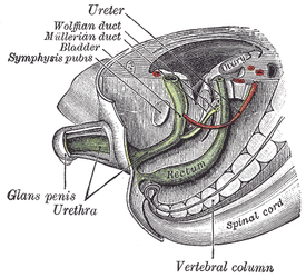
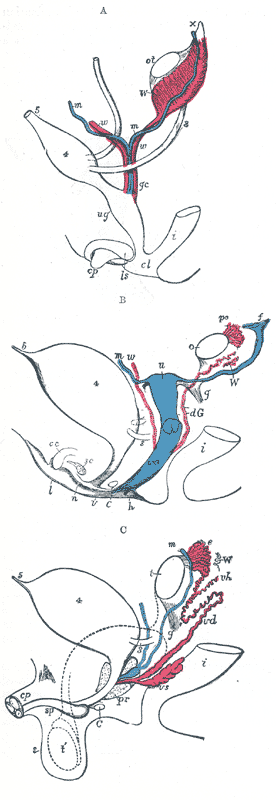
Internal organs
| Embryological precursor | Female | Male |
|---|---|---|
| 3. Ureter | Ureter | Ureter |
| 4. Urinary bladder | Urinary bladder | Urinary bladder |
| 5. Urachus | Urachus | Urachus |
| i. Lower part of the intestine | i. Lower part of the intestine | i. Lower part of the intestine |
| cl. Cloaca | ||
| cp. Elevation which becomes clitoris or penis (genital tubercle) | cc. Corpus cavernosum clitoridis | cp. Corpora cavernosa penis cut short |
| ug. Sinus urogenitalis | C. Greater vestibular gland, and immediately above it the urethra | C. Bulbo-urethral gland of one side |
| f. The abdominal opening of the left uterine tube | ||
| g. Round ligament, corresponding to gubernaculum | g. The gubernaculum | |
| h. Situation of the hymen | ||
| ls. Labioscrotal folds | l. Labium majus | s. Scrotum |
| n. Labium minus | ||
| m, m. Right and left Müllerian ducts uniting together and running with the Wolffian ducts in gc, the genital cord | m. Müllerian duct, the upper part of which remains as the hydatid of Morgagni; the lower part, represented by a dotted line descending to the prostatic utricle, constitutes the occasionally existing cornu and tube of the uterus masculinus | |
| ot. The genital ridge from which either the ovary or testis is formed. | o. The left ovary | t. Testis in the place of its original formation; t’, together with the dotted lines above, indicates the direction in which the testis and epididymis descend from the abdomen into the scrotum. |
| pr. The prostate | ||
| sc. Corpus cavernosum urethrae | sp. Corpus cavernosum urethrae | |
| u. Uterus. The uterine tube of the right side is marked m. | ||
| v. Vulva | ||
| va. Vagina | ||
| vh. Ductus aberrans | ||
| vs. The vesicula seminalis | ||
| W. Left Wolffian body | W. Scattered remains of the Wolffian body, constituting the organ of Giraldès, or the paradidymis of Waldeyer. | |
| w, w. Right and left Wolffian ducts | W. Scattered remains of Wolffian tubes near it (paroöphoron of Waldeyer); dG. Remains of the left Wolffian duct, such as give rise to the duct of Gärtner, represented by dotted lines; that of the right side is marked w. | |
| po. Epoophoron |
External organs
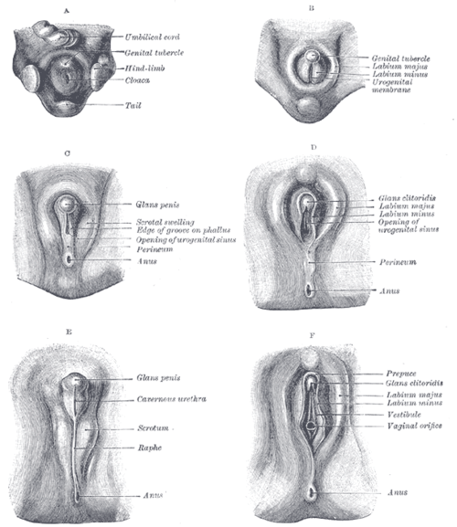
- A: Undifferentiated
- B: Female
- C: Male
- D: Female
- E: Male
- F: Female
The external genitalia of both males and females have similar origins. They arise from the genital tubercle that forms anterior to the cloacal folds (proliferating mesenchymal cells around the cloacal membrane). The caudal aspect of the cloacal folds further subdivides into the posterior anal folds and the anterior urethral folds. Bilateral to the urethral fold, genital swellings (tubercles) become prominent. These structures are the future scrotal swellings and labia majora in males and females, respectively.
The genital tubercles of an eight week old embryo of either sex are identical. They both have a glans area, which will go on to form the glans clitoridis (females) or glans penis (males), a urogenital fold and groove, and an anal tubercle. At around ten weeks, the external genitalia are still similar. At the base of the glans, there is a groove known as the coronal sulcus or corona glandis. It is the site of attachment of the future prepuce. Just anterior to the anal tubercle, the caudal end of the left and right urethral folds fuse to form the urethral raphe. The lateral part of the genital tubercle (called the lateral tubercle) grows longitudinally and is about the same length in either sex.
Human physiology
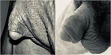
The male external genitalia include the penis, the male urethra, and the scrotum. The female external genitalia include the clitoris, the labia majora, and the labia minora, which are collectively called the vulva. External genitalia vary widely in external appearance among different people.
Most scientists and scholars agree that the glans clitoridis and the glans penis each contain some 7,000 sensory nerve endings. There was a 1999 study in which a group of scientists claimed the clitoris had more nerve endings than the penis;[2] however, this has been disproven with more recent studies. The difference is that the glans clitoridis packs them into a volume only about one-tenth the size of the glans penis. In addition, the glans clitoridis has greater variability in cutaneous corpuscular receptor density (1-14 per 100× high-powered field) compared with the glans penis (1-3 per 100× high-power field). Touch for touch, this concentration of nerves makes the glans clitoridis more sensitive than the glans penis. As a result, many women can feel discomfort—even pain—with anything more than a gentle touch.[3]
References
- Cai Y (2009). "Revisiting old vaginal topics: conversion of the Müllerian vagina and origin of the "sinus" vagina". Int J Dev Biol. 53 (7): 925–34. doi:10.1387/ijdb.082846yc. PMID 19598112.
- Angier, Natalie. “Woman: An Intimate Geography“, Houghton Mifflin Company, New York, 1999. ISBN 0395691303.
- Cheryl Shih, Christopher J. Cold, and Claire C. Yang. “Cutaneous Corpuscular Receptors of the Human Glans Clitoris: Descriptive Characteristics and Comparison with the Glans Penis“, Journal of Sexual Medicine, Elsevier, July 2013.
This article incorporates text in the public domain from the 20th edition of Gray's Anatomy (1918)