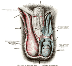
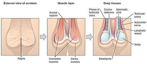
Scrotum
The scrotum is an anatomical male reproductive structure that consists of a suspended dual-chambered sack of skin and smooth muscle that is present in most terrestrial male mammals and located under the penis. One testis is typically lower than the other to avoid compression in the event of impact.[1] The perineal raphe is a small, vertical, slightly raised ridge of scrotal skin under which is found the scrotal septum. It appears as a thin longitudinal line that runs front to back over the entire scrotum. The scrotum contains the external spermatic fascia, testes, epididymis, and ductus deferens. It is a distention of the perineum and carries some abdominal tissues into its cavity including the testicular artery, testicular vein, and pampiniform plexus. In humans and some other mammals, the scrotum becomes covered with pubic hair at puberty. The scrotum will usually tighten during penile erection and when exposed to cold temperature.
| Scrotum | |
|---|---|
 Human scrotum in a relaxed state (left) and a tense state (right) | |
| Details | |
| Precursor | Labioscrotal folds |
| Artery | Anterior scrotal artery & Posterior scrotal artery |
| Vein | Testicular vein |
| Nerve | Posterior scrotal nerves, Anterior scrotal nerves, genital branch of genitofemoral nerve, perineal branches of posterior femoral cutaneous nerve |
| Lymph | Superficial inguinal lymph nodes |
| Identifiers | |
| Latin | Scrotum |
| MeSH | D012611 |
| TA | A09.4.03.001 |
| FMA | 18252 |
| Anatomical terminology | |
The scrotum is biologically homologous to the labia majora in females. Although present in most mammals, the external scrotum is absent in streamlined marine mammals, such as whales and seals,[2] as well as in some lineages of land mammals, such as the afrotherians, xenarthrans, and numerous families of bats, rodents, and insectivores.[3][4]
Structure
Nerve supply
| Nerve | Surface[5] |
|---|---|
| Genital branch of genitofemoral nerve | anterolateral |
| Anterior scrotal nerves (from ilioinguinal nerve) | anterior |
| Posterior scrotal nerves (from perineal nerve) | posterior |
| perineal branches of posterior femoral cutaneous nerve | inferior |
Blood supply
| Blood vessels[6] | |
|---|---|
| Anterior scrotal artery | originates from the deep external pudendal artery[7] |
| Posterior scrotal artery | |
| Testicular artery | |
Skin and glands
| Skin associated tissues [6] | |
|---|---|
| Pubic hair | |
| Sebaceous glands | |
| Apocrine glands | |
| Smooth muscle | |
The skin on the scrotum is more highly pigmented compared to the rest of the body. The septum is a connective tissue membrane dividing the scrotum into two cavities. [8]
Lymphatic system
The scrotum lymph drains initially into the superficial inguinal lymph nodes, this then drains into the deep inguinal lymph nodes. The deep inguinal lymph nodes drain into the common iliac which ultimately releases lymph into the cisterna chyli.
| Lymphatic vessels[9] | |
|---|---|
| Superficial inguinal lymph nodes | |
| Popliteal lymph nodes | |
Asymmetry
One testis is typically lower than the other, which is believed to function to avoid compression in the event of impact; in humans, the left testis is typically lower than the right.[1] An alternative view is that testis descent asymmetry evolved to enable more effective cooling of the testicles.[10]
Internal structure
Additional tissues and organs reside inside the scrotum and are described in more detail in the following articles:
- Appendix of epididymidis
- Cavity of tunical albuginea
- Cremaster muscle
- Dartos
- Ductus Deferens
- Efferent ductules
- Epididymis
- Leydig cell
- Lobule of testes
- Paradidymis
- Rete testes
- Scrotal septum
- Seminiferous tubule
- Sertoli cell
- Spermatic cord
- Testes
- Tunica albuginea of testis
- Tunica vaginalis parietal layer
- Tunica vaginalis visceral layer
- Tunica vasculosa testis
- Vas deferens
Development
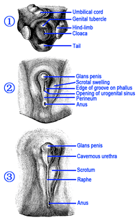
Genital homology between sexes
Male sex hormones are secreted by the testes later in embryonic life to cause the development of secondary sex organs. The scrotum is developmentally homologous to the labia majora. The raphe does not exist in females. Reproductive organs and tissues develop in females and males begin during the fifth week after fertilization. The gonadal ridge grows behind the peritoneal membrane. By the sixth week, string-like tissues called primary sex cords form within the enlarging gonadal ridge. Externally, a swelling called the genital tubercule appears over the cloacal membrane.
Up until the eighth week after fertilization, the reproductive organs do not appear to be different between the male and female and are called in-differentiated. Testosterone secretion starts during week eight, reaches peak levels during week 13 and eventually declines to very low levels by the end of the second trimester. The testosterone causes the masculinization of the labioscrotal folds into the scrotum. The scrotal raphe is formed when the embryonic, urethral groove closes by week 12.[11]
Function
The scrotum regulates the temperature of the testes and maintains it at 35 degrees Celsius (95 degrees Fahrenheit), i.e. two degrees below the body temperature of 37 degrees Celsius (98.6 degrees Fahrenheit). Higher temperatures affect spermatogenesis[13] Temperature control is accomplished by the smooth muscles of the scrotum moving the testicles either closer to or further away from the abdomen dependent upon the ambient temperature. This is accomplished by the cremaster muscle in the abdomen and the dartos fascia (muscular tissue under the skin).[12]
Having the scrotum and testicles situated outside the abdominal cavity may provide additional advantages. The external scrotum is not affected by abdominal pressure. This may prevent the emptying of the testes before the sperm were matured sufficiently for fertilization.[13] Another advantage is it protects the testes from jolts and compressions associated with an active lifestyle. Animals that move at a steady pace – such as elephants, whales, and marsupial moles – have internal testes and no scrotum.[14] Unlike placental mammals, some male marsupials have a scrotum that is anterior to the penis,[15][16][17] although there are several marsupial species without an external scrotum.[18] In humans, the scrotum may provide some friction during intercourse, helping to enhance the activity.[19]
Clinical significance
A study has indicated that use of a laptop computer positioned on the lap can negatively affect sperm production.[20][21]
Diseases and conditions
The scrotum and its contents can develop diseases or incur injuries. These include:
- Candidiasis (yeast infection)
- sebaceous cyst
- epidermal cyst
- hydrocele
- hematocele
- Molluscum contagiosum
- spermatocele
- Paget's disease of the scrotum[22]
- varicocele
- inguinal hernia
- epididymo-orchitis
- testicular torsion
- genital warts
- testicular cancer
- dermatitis
- undescended testes
- Chyloderma
- mumps
- scabies
- herpes
- pubic lice
- Chancroid (Haemophilus ducreyi)
- Chlamydia (Chlamydia trachomatis)
- Gonorrhea (Neisseria gonorrhoeae)
- Granuloma inguinale or (Klebsiella granulomatis)
- Syphilis (Treponema pallidum)
- scrotum eczema
- scrotal psoriasis disease
- Riboflavin deficiency
- Chimney sweeps' carcinoma[23]
See also
- Scrotal infusion, a temporary form of body modification
- Sex organ
- Retroperitoneal lymph node dissection
- Testicular self-examination
Bibliography
- Books
- This article incorporates text in the public domain from page 1237 of the 20th edition of Gray's Anatomy (1918)
- Van De Graaff, Kent M.; Fox, Stuart Ira (1989). Concepts of Human Anatomy and Physiology. Dubuque, Iowa: William C. Brown Publishers. ISBN 978-0697056757.
- Elson, Lawrence; Kapit, Wynn (1977). The Anatomy Coloring. New York, New York: Harper & Row. ISBN 978-0064539142.
- "Gross Anatomy Image". Medical Gross Anatomy Atlas Images. University of Michigan Medical School. 1997. Retrieved 2015-02-23.
- Berkow, MD, editor, Robert (1977). The Merck Manual of Medical Information; Home Edition. Whitehouse Station, New Jersey: Merck Research Laboratories. ISBN 978-0911910872.CS1 maint: multiple names: authors list (link) CS1 maint: extra text: authors list (link)
References
| Wikimedia Commons has media related to Scrotums. |
- Anthony F.Bogaert, "Genital asymmetry in men Archived 2015-05-28 at the Wayback Machine", Human Reproduction vol.12 no.1 pp.68–72, 1997. PMID 9043905.
- William F. Perrin; Bernd Würsig; J.G.M. Thewissen (26 February 2009). Encyclopedia of Marine Mammals. Academic Press. ISBN 978-0-08-091993-5.
- "Scrotum". National Institutes of Health. Retrieved 6 January 2011.
- Lovegrove, B. G. (2014). "Cool sperm: Why some placental mammals have a scrotum". Journal of Evolutionary Biology. 27 (5): 801–814. doi:10.1111/jeb.12373. PMID 24735476.
- Moore, Keith; Anne Agur (2007). Essential Clinical Anatomy, Third Edition. Lippincott Williams & Wilkins. p. 132. ISBN 978-0-7817-6274-8.
- Elson 1977.
- antthigh at The Anatomy Lesson by Wesley Norman (Georgetown University)
- "Scrotum". Encyclopædia Britannica. Retrieved 2015-02-24.
- "VIII. The Lymphatic System. 5. The Lymphatics of the Lower Extremity. Gray, Henry. 1918. Anatomy of the Human Body". Retrieved 2015-02-24.
- Gallup, Gordon G.; Finn, Mary M.; Sammis, Becky (2009). "On the Origin of Descended Scrotal Testicles: The Activation Hypothesis". Evolutionary Psychology. 7 (4): 147470490900700. doi:10.1177/147470490900700402.
- Van de Graaff 1989, p. 927-931.
- Van de Graaff 1989, p. 935.
- Van de Graaff 1989, p. 936.
- "Science : Bumpy lifestyle led to external testes - 17 August 1996 - New Scientist". New Scientist. Retrieved 2007-11-06.
- Hugh Tyndale-Biscoe; Marilyn Renfree (30 January 1987). Reproductive Physiology of Marsupials. Cambridge University Press. ISBN 978-0-521-33792-2.
- Libbie Henrietta Hyman (15 September 1992). Hyman's Comparative Vertebrate Anatomy. University of Chicago Press. pp. 583–. ISBN 978-0-226-87013-7.
- Menna Jones; Chris R. Dickman; Michael Archer (2003). Predators with Pouches: The Biology of Carnivorous Marsupials. Csiro Publishing. ISBN 978-0-643-06634-2.
- C. Hugh Tyndale-Biscoe (2005). Life of Marsupials. Csiro Publishing. ISBN 978-0-643-06257-3.
- Jones, Richard (2013). Human Reproductive Biology. Academic Press. p. 74. ISBN 9780123821850.
The rear-entry position of mating may allow the scrotum to stimulate the clitoris and, in this way, may produce an orgasm ...
- "Laptops may damage male fertility". BBC News. 2004-12-09. Retrieved 2012-01-30.
- Sheynkin, Yefim; et al. (February 2005). "Increase in scrotal temperature in laptop computer users". Hum. Reprod. 20 (2): 452–455. doi:10.1093/humrep/deh616. PMID 15591087.
- "Paget's disease of the scrotum Symptoms, Diagnosis, Treatments and Causes". RightDiagnosis.com. Retrieved 2015-02-24.
- TCMwell.
Outline of human sexuality | |
|---|---|
| Physiology and biology |
|
| Health and education |
|
| Identity and diversity |
|
| Law |
|
| History |
|
| Relationships and society |
|
| By country |
|
| Sexual activities |
|
| Sex industry |
|
| Religion and sexuality |
|
| |
| Authority control |
|
|---|