

Anaplasmosis
 ShareCompartir
Agent
Anaplasma phagocytophilum
 WHERE FOUND
Anaplasmosis is most frequently reported from the upper midwest and northeastern U.S. in areas that correspond with the known geographic distribution of Lyme disease.
INCUBATION PERIOD: 1-2 weeks
SIGNS AND SYPMTOMS
- Fever, shaking, chills
- Severe headache
- Malaise
- Myalgia
- Gastrointestinal symptoms (nausea, vomiting, diarrhea, anorexia)
- Cough
- Rash (rare cases)
Anaplasmosis and ehrlichiosis have similar clinical presentations, but they are transmitted by two different species of ticks and generally occur in different regions of the U.S.
Anaplasmosis was formerly known as Human Granulocytic Ehrlichiosis (HGE).
The signs and symptoms list presents symptoms commonly seen with anaplasmosis. However, it is important to note that few people will develop all symptoms and the number and combination of symptoms varies greatly from person to person.
 Confirmation of the diagnosis is based on laboratory testing, but antibiotic therapy should not be delayed in a patient with a suggestive clinical presentation.
GENERAL LABORATORY FINDINGS
Typically Observed During the First Week of Clinical Disease:
- Mild anemia
- Thrombocytopenia
- Leukopenia (characterized by relative and absolute lymphopenia and a left shift)
- Mild to moderate elevations in hepatic transaminases may occur in some patients.
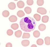
- Visualization of morulae in the cytoplasm of granulocytes during examination of blood smears is highly suggestive of a diagnosis; however, blood smear examination is insensitive and should never be relied upon solely to rule anaplasmosis in or out.
LABORATORY CONFIRMATION
Antibodies to A. phagocytophilum are detectable 7–10 days after illness onset. The gold-standard serologic test looks for a four-fold change in antibody titers using indirect immunofluorescence assay (IFA) on paired samples. The first sample should be taken within the first week of illness and the second should be taken 2 to 4 weeks later.
- Demonstration of a four-fold change in IgG-specific antibody titer by indirect immunofluorescence assay (IFA) test in paired serum samples; or
- Detection of DNA by PCR of whole blood. This method is most sensitive within the first week of illness; sensitivity may decrease after administration of antibiotics.
NOTE: IgM antibodies are less specific than IgG antibodies and are more likely to generate false positives. IgM results alone should not be used for laboratory diagnosis.
NOTE: Consider the possibility of coinfection with Babesia microti and/or Borrelia burgdorferi.
NOTE: Antibody titers are frequently negative in the first 7–10 days of illness, thus serologic tests may be falsely negative during this time period.
Treatment
Anaplasmosis, ehrlichiosis, and Rocky Mountain spotted fever are treated in the same manner with doxycycline.† Clinical suspicion of any of these diseases is sufficient to begin treatment. Delay in treatment may result in severe illness and even death. The regimens listed below are guidelines only and may need to be adjusted depending on a patient’s age, medical history, underlying health conditions, pregnancy status, or allergies. Consult an infectious disease specialist in cases of documented pregnancy or life-threatening allergies to doxycycline.
| Age Category | Drug | Dosage | Maximum | Duration (Days) |
|---|---|---|---|---|
| Adults | Doxycycline | 100 mg twice per day, orally or IV | 100 mg/dose | Patients with suspected anaplasmosis infection should be treated with doxycycline for 10-14 days to provide appropriate length of therapy for possible incubating co-infection with Lyme disease |
| Children weighing <100 lbs. (45.4 kg) | Doxycycline | 2.2 mg/kg per dose twice per day, orally or IV | 100 mg/dose |
NOTE: Use doxycycline as first-line treatment for suspected anaplasmosis in patients of all ages. The use of doxycycline to treat suspected anaplasmosis in children is recommended by both the CDC and the American Academy of Pediatrics Committee on Infectious Diseases. Use of antibiotics other than doxycycline increases the risk of patient death. At the recommended dose and duration needed to treat anaplasmosis, no evidence has been shown to cause staining of permanent teeth, even when five courses are given before the age of eight.
References
Bakken JS, Aguero-Rosenfeld ME, Tilden RL, et al. Serial measurements of hematologic counts during the active phase of human granulocytic ehrlichiosis. Clin Infect Dis 2001; 32: 862–870.
†Biggs HM, Barton Behravesh C, Bradley KK, et al. Diagnosis and management of tickborne rickettsial diseases: Rocky Mountain spotted fever and other spotted fever group rickettsioses, ehrlichioses, and anaplasmosis — United States. MMWR Recomm Rep 2016; 65 (2); 1-48.
Engel J, Bradley K, et al. Revision of the national surveillance case definition for ehrlichiosis (ehrlichiosis/anaplasmosis). Council of State and Territorial Epidemiologists, Infectious Diseases Committee, 2007 Position Statement. [PDF – 36 KB]
Gelfand JA, Vannier E. Ehrlichia chaffeensis (human monocytotropic ehrlichiosis), Anaplasma phagocytophilum (human granulocytotropic anaplasmosis) and other ehrlichiae. In: Mandell GL, Bennett JE, Dolin R, editors. Mandell, Douglas, and Bennett’s Principles and Practice of Infectious Diseases. 6th ed. Philadelphia, PA: Churchill Livingstone; 2005. p. 2310–2318.
Todd SR, Dahlgren FS, et al. No visible dental staining in children treated with doxycycline for suspected Rocky Mountain spotted fever. J Pediatr, available online 17 March 2015.
Wormser GP, Dattwyler RJ, Shapiro ED, et al. The clinical assessment, treatment and prevention of Lyme disease, human granulocytic anaplasmosis, and babesiosis: clinical practice guidelines by the Infectious Diseases Society of America. Clin Infect Dis 2006; 43:1089–1134.
Reported Cases of Anaplasmosis, U.S., 2015
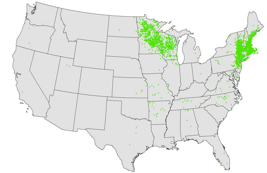
NOTE: Each dot represents one case. Cases are reported from the infected person’s county of residence, not necessarily the place where they were infected.
NOTE: In 2015, no cases of anaplasmosis were reported from Hawaii or Alaska.
- Page last reviewed: October 23, 2014
- Page last updated: February 8, 2017
- Content source: