Vorticose veins
The vorticose veins, referred to clinically as the vortex veins, drain the ocular choroid. The number of vortex veins is known to vary from 4 to 8 with about 65% of the normal population having 4 or 5.[1] In most cases, there is at least one vortex vein in each quadrant. Typically, the entrances to the vortex veins in the outer layer of the choroid (lamina vasculosa) can be observed funduscopically and provide an important clinical landmarks identifying the ocular equator. However, the veins run posteriorly in the sclera exiting the eye well posterior to the equator.
| Vorticose veins | |
|---|---|
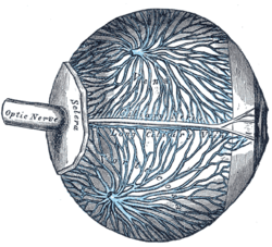 The veins of the choroid. (Venae vorticosae labeled - though difficult to see - at center.) | |
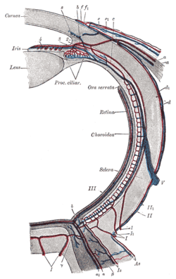 Diagram of the blood vessels of the eye, as seen in a horizontal section. ("V", at center right, is the label for the vena vorticosa) | |
| Details | |
| Drains to | inferior ophthalmic vein, superior ophthalmic vein |
| Artery | short posterior ciliary arteries |
| Identifiers | |
| Latin | venae vorticosae |
| TA | A12.3.06.106 |
| FMA | 70880 |
| Anatomical terminology | |
Some vortex veins drain into the superior ophthalmic vein which drains into the cavernous sinus.[2][3] Some vortex veins drain into the inferior ophthalmic vein which drains into the pterygoid plexus and cavernous sinus.[2][4] There is usually collateral circulation between the superior and inferior orbital veins.[5][6][2][7]
Additional images
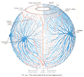 The blood-vessels of the eyeball (diagrammatic).
References
This article incorporates text in the public domain from page 1010 of the 20th edition of Gray's Anatomy (1918)
- Kutoglu, T., Yalcin, B., Kocabiyik, N. and Ozan, H. (2005), Vortex veins: Anatomic investigations on human eyes. Clinical Anatomy, 18: 269–273. doi:10.1002/ca.20092
- Drake, Richard (2020). Gray's anatomy for students. Philadelphia, PA: Elsevier. ISBN 978-0-323-39304-1. OCLC 1089399265.
- "The VisionHelp Blog". WordPress. 2019-06-28. Retrieved 2019-06-28.
- Remington, Lee Ann (2012). "Orbital Blood Supply". Clinical Anatomy and Physiology of the Visual System. Elsevier. pp. 202–217. doi:10.1016/b978-1-4377-1926-0.10011-6. ISBN 978-1-4377-1926-0.
- "Dilated superior ophthalmic vein commonly caused by cerebral vascular malformation". American Academy of Ophthalmology. 2018-05-07. Retrieved 2019-06-28.
- Christopher J Murphy
- BASIC HUMAN ANATOMY - O'RAHILLY, MÜLLER, CARPENTER & SWENSON