Great cerebral vein
The great cerebral vein is one of the large blood vessels in the skull draining the cerebrum of the brain. It is also known as the "vein of Galen", named for its discoverer, the Greek physician Galen. However, it is not the only vein with this eponym.
| Great cerebral vein | |
|---|---|
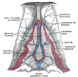 Velum interpositum. (Great cerebral vein labeled at bottom center.) | |
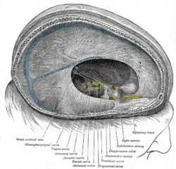 Dura mater and its processes exposed by removing part of the right half of the skull, and the brain. (Great cerebral vein labeled at bottom left.) | |
| Details | |
| Drains from | cerebrum |
| Source | internal cerebral veins |
| Drains to | straight sinus |
| Artery | cerebral arteries |
| Identifiers | |
| Latin | vena magna cerebri |
| TA | A12.3.06.027 |
| FMA | 50993 |
| Anatomical terminology | |
Structure
The great cerebral vein is considered as one of the deep cerebral veins. Other deep cerebral veins are the internal cerebral veins, formed by the union of the superior thalamostriate vein and the superior choroid vein at the interventricular foramina. The internal cerebral veins can be seen on the superior surfaces of the caudate nuclei and thalami just under the corpus callosum.[1] The veins at the anterior poles of the thalami merge posterior to the pineal gland to form the great cerebral vein.[1] Most of the blood in the deep cerebral veins collects into the great cerebral vein.[2] This comes from the inferior side of the posterior end of the corpus callosum and empties ie similarities, there are also differences between these two types of veins in the brain. The superficial veins at the dorsal parts of the hemispheres run upward and medially and empty into the large superior sagittal sinus in the upper margin of the falx cerebri. The superior sagittal sinus divides into two parts called the transverse sinuses where the falx cerebri meets the tentorium cerebelli.[3] The sigmoid sinus, which continues the transverse sinus, empties into the jugular vein at the jugular foramen. The internal jugular vein leaves the skull and travels downward to the neck.[3]
The veins of the brain have very thin walls and contain no valves. They emerge in the brain and lie in the subarachnoid space. They pierce the arachnoid matter and the meningeal layer in the dura and drain into the cranial venous sinuses.[3]
Clinical significance
Malformations
Most conditions associated with the great cerebral vein are due to congenital defects. Vein of Galen aneurysmal malformations (VGAM) are the most common form of symptomatic cerebrovascular malformation in neonates and infants.[4] The presence and locations of angiomas are very variable and do not follow any predictable pattern.[5] The congenital malformation develops during weeks 6-11 of fetal development as a persistent embryonic prosencephalic vein of Markowski; thus, VGAM is actually a misnomer. The vein of Markowski actually drains into the vein of Galen.
Absence
Absence of the great cerebral vein is a congenital disorder. The deep cerebral veins of the brain normally drain through the great cerebral vein. In its absence, the veins from the diencephalon and the basal ganglia drain laterally into the transverse sinus instead of conjoining in the midline through the Galenic drainage system.[6] Absence of the great cerebral vein is quite rare. It is detected in infancy and most patients die in the neonatal period or in early infancy.
Thrombosis
Thrombosis of the great cerebral vein is a form of stroke due to a blood clot in the vein. It affects just 3 to 8% of patients, predominantly women.[7] Patients may present with consciousness problems, headaches, nausea, visual defects, fatigue, disturbance of eye movements and pupillary reflexes, or coma.[7] Thrombosis of the cerebral vein is often deadly but can be survived. Risk factors include oral contraceptives, pregnancy, and the postpartum period.[7]
History
See also
References
- Diamond, Marian C.; Elson, Lawrence M.; Scheibel, Arnold B. (1985). "Venous Drainage of the Cerebral Hemispheres". The Human Brain Coloring Book. HarperCollins. ISBN 978-0-06-460306-5.
- Brodal, Per (2004) [1992]. The central nervous system: structure and function (3rd (rev) ed.). Oxford University Press. pp. 100–102. ISBN 978-0-19-516560-9.
- Snell, Richard S. (August 1997) [1980]. Clinical Neuroanatomy: A Review with Questions and Explanations (2nd ed.). Philadelphia: Lippincott Williams & Wilkins. pp. 217–219. ISBN 978-0-316-80315-1.
- Johnston IH; Whittle IR; Besser M; Morgan MK (May 1987). "Vein of Galen malformation: diagnosis and management". Neurosurgery. 20 (5): 747–758. doi:10.1227/00006123-198705000-00013. ISSN 0148-396X. PMID 3601022.
- Vidyasagar C (April 2005). "Persistent embryonic veins in the arteriovenous malformation of the diencephalon". Acta Neurochirurgica. 47 (1): 63–82. doi:10.1007/BF01404664. ISSN 0942-0940. PMID 474205.
- Lasjaunias P; Garcia-Monaco R; Rodesch G; Terbrugge K (May 1991). "Deep venous drainage in great cerebral vein (vein of Galen) absence and malformations". Neuroradiology. 33 (3): 234–238. doi:10.1007/BF00588224. ISSN 1432-1920. PMID 1881541.
- van den Bergh WM; van der Schaaf I; van Gijn J (July 2005). "The spectrum of presentations of venous infarction caused by deep cerebral vein thrombosis". Neurology. 65 (2): 192–196. doi:10.1212/01.wnl.0000179677.84785.63. ISSN 1526-632X. PMID 16043785.
External links
- "Anatomy diagram: 13048.000-3". Roche Lexicon - illustrated navigator. Elsevier. Archived from the original on 2014-01-01.
- http://neuroangio.org/venous-brain-anatomy/deep-venous-system/
- Vein of Galen images
| Authority control |
|---|