Superficial temporal vein
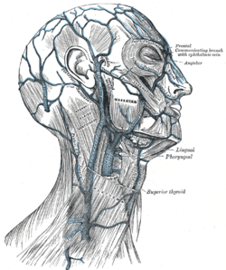
The superficial temporal vein is a vein of the side of the head. It begins on the side and vertex of the skull in a network of veins which communicates with the frontal vein and supraorbital vein, with the corresponding vein of the opposite side, and with the posterior auricular vein and occipital vein. It ultimately crosses the posterior root of the zygomatic arch, enters the parotid gland, and unites with the internal maxillary vein to form the posterior facial vein.
| Superficial temporal vein | |
|---|---|
 Veins of the head and neck. ("Sup. Temp." labeled at center, anterior to the ear.) | |
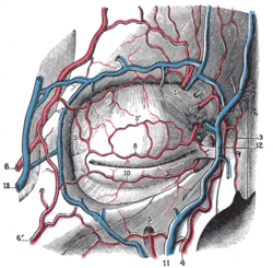
 Bloodvessels of the eyelids, front view. (13, at left, is branch of the superficial temporal vein.) | |
| Details | |
| Drains from | temple, scalp |
| Drains to | retromandibular vein |
| Artery | superficial temporal artery |
| Identifiers | |
| Latin | venae temporales superficiales |
| TA | A12.3.05.032 |
| FMA | 70849 |
| Anatomical terminology | |
Structure
It begins on the side and vertex of the skull in a network (plexus) which communicates with the frontal vein and supraorbital vein, with the corresponding vein of the opposite side, and with the posterior auricular vein and occipital vein.
From this network frontal and parietal branches arise, and join above the zygomatic arch to form the trunk of the vein, which is joined by the middle temporal vein emerging from the temporalis muscle.
It then crosses the posterior root of the zygomatic arch, enters the substance of the parotid gland, and unites with the internal maxillary vein to form the posterior facial vein.
Tributaries
The superficial temporal vein receives in its course the following:
- some parotid veins
- articular veins from the temporomandibular joint
- anterior auricular veins from the auricula
- the transverse facial vein from the side of the face
References
This article incorporates text in the public domain from page 645 of the 20th edition of Gray's Anatomy (1918)
External links
- lesson4 at The Anatomy Lesson by Wesley Norman (Georgetown University) (parotid2)
- Diagram at Tufts.edu
- Diagram at stchas.edu