Uterine gland
Uterine glands or endometrial glands are tubular glands, lined by ciliated columnar epithelium, found in the functional layer of the endometrium that lines the uterus.[1] Their appearance varies during the menstrual cycle. During the proliferative phase, uterine glands appear long due to estrogen secretion by the ovaries. During the secretory phase, the uterine glands become very coiled with wide lumens and produce a glycogen-rich secretion. This change corresponds with an increase in blood flow to spiral arteries due to increased progesterone secretion from the corpus luteum. During the pre-menstrual phase, progesterone secretion decreases as the corpus luteum degenerates, which results in decreased blood flow to the spiral arteries. The functional layer of the uterus containing the glands becomes necrotic, and eventually sloughs off during the menstrual phase of the cycle.
| Uterine glands | |
|---|---|
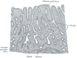 Vertical section of mucous membrane of human uterus. (Glands labeled at center right.) | |
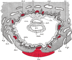 Section through ovum imbedded in the uterine decidua. Semidiagrammatic. am. Amniotic cavity. b.c. Blood-clot. b.s. Body-stalk. ect. Embryonic ectoderm. ent. Entoderm. mes. Mesoderm. m.v. Maternal vessels. tr. Trophoblast. u.e. Uterine epithelium. u.g. Uterine glands. y.s. Yolk-sac. | |
| Details | |
| Identifiers | |
| Latin | glandulae uterinae |
| TA | A09.1.03.028 |
| FMA | 71647 |
| Anatomical terminology | |
They are of small size in the unimpregnated uterus, but shortly after impregnation become enlarged and elongated, presenting a contorted or waved appearance.
Function
The uterine glands synthesize or transport and secrete substances essential for survival and development of the embryo or fetus and associated extraembryonic membranes.[2]
Some secretory components from the uterine glands are taken up by the secondary yolk sac lining the exocoelomic cavity during pregnancy, and may thereby assist in providing fetal nutrition.[3]
Additional images
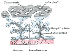 Primary chorionic villi. Diagrammatic.
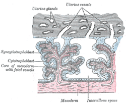 Secondary chorionic villi. Diagrammatic.
See also
References
This article incorporates text in the public domain from page 1262 of the 20th edition of Gray's Anatomy (1918)
- "Uterine Gland - Embryology". embryology.med.unsw.edu.au.
- Gray CA, Bartol FF, Tarleton BJ, et al. (November 2001). "Developmental biology of uterine glands". Biol. Reprod. 65 (5): 1311–23. doi:10.1095/biolreprod65.5.1311. PMID 11673245.
- Burton GJ, Watson AL, Hempstock J, Skepper JN, Jauniaux E (June 2002). "Uterine glands provide histiotrophic nutrition for the human fetus during the first trimester of pregnancy". J. Clin. Endocrinol. Metab. 87 (6): 2954–9. doi:10.1210/jc.87.6.2954. PMID 12050279.
External links
- Swiss embryology (from UL, UB, and UF) gnidation/role02
- Histology at KUMC epithel-epith06
- Anatomy photo: Reproductive/mammal/uterus1/uterus2 - Comparative Organology at University of California, Davis - "Mammal, uterus (LM, Medium)"
- UIUC Histology Subject 1024
| Authority control |
|---|