Purkinje fibers
The Purkinje fibers (/pɜːrˈkɪndʒi/ pur-KIN-jee;[1] Purkinje tissue or subendocardial branches) are located in the inner ventricular walls of the heart, just beneath the endocardium in a space called the subendocardium. The Purkinje fibers are specialised conducting fibers composed of electrically excitable cells that are larger than cardiomyocytes with fewer myofibrils and many mitochondria and which (cells) conduct cardiac action potentials more quickly and efficiently than any other cells in the heart.[2] Purkinje fibers allow the heart's conduction system to create synchronized contractions of its ventricles, and are, therefore, essential for maintaining a consistent heart rhythm.
| Purkinje fibers | |
|---|---|
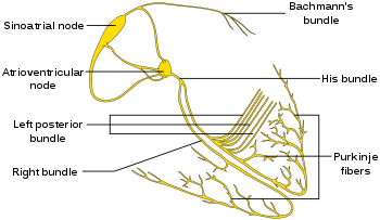 Isolated heart conduction system showing Purkinje fibers | |
 The QRS complex is the large peak. | |
| Details | |
| Identifiers | |
| Latin | Rami subendocardiales |
| MeSH | D011690 |
| TA | A12.1.06.008 |
| FMA | 9492 |
| Anatomical terminology | |
Histology
Purkinje fibers are a unique cardiac end-organ. Further histologic examination reveals that these fibers are split in ventricles walls. The electrical origin of atrial Purkinje fibers arrives from the sinoatrial node.
Given no aberrant channels, the Purkinje fibers are distinctly shielded from each other by collagen or the cardiac skeleton.
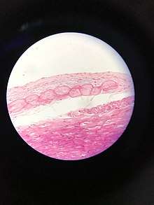
The Purkinje fibers are further specialized to rapidly conduct impulses (numerous fast voltage-gated sodium channels and mitochondria, fewer myofibrils than the surrounding muscle tissue). Purkinje fibers take up stain differently from the surrounding muscle cells because of relatively fewer myofibrils than other cardiac cells and the presence of glycogen around the nucleus causes Purkinje fibers to appear, on a slide, lighter and larger than their neighbors, arranged along the longitudinal direction (parallel to the cardiac vector). They are often binucleated cells.
Function
Heart rate is governed by many influences from the autonomic nervous system. The Purkinje fibers do not have any known role in setting heart rate unless the SA node is compromised. They are influenced by electrical discharge from the sinoatrial node.
During the ventricular contraction portion of the cardiac cycle, the Purkinje fibers carry the contraction impulse from both the left and right bundle branch to the myocardium of the ventricles. This causes the muscle tissue of the ventricles to contract and generate force to eject blood out of the heart, either to the pulmonary circulation from the right ventricle or to the systemic circulation from the left ventricle.[3]
Purkinje fibers also have the ability of firing at a rate of 15-40 beats per minute if upstream conduction or pacemaking ability is compromised. In contrast, the SA node in normal state can fire at 60-100 beats per minute. In short, they generate action potentials, but at a slower rate than sinoatrial node. This capability is normally suppressed. Thus, they serve as the last resort when other pacemakers fail. When a Purkinje fiber does fire, it is called a premature ventricular contraction or PVC, or in other situations can be a ventricular escape. It plays a vital role in the circulatory system.
Etymology
They are named after Jan Evangelista Purkyně who discovered them in 1839.
References
- Jones, Daniel (2011). Roach, Peter; Setter, Jane; Esling, John (eds.). Cambridge English Pronouncing Dictionary (18th ed.). Cambridge University Press. ISBN 978-0-521-15255-6.
- "Purkinje fiber." The American Heritage® Medical Dictionary. 2007. Houghton Mifflin Company 23 Oct. 2016 http://medical-dictionary.thefreedictionary.com/Purkinje+fiber
- Podrid, Philip J.; Kowey, Peter R. (2010). Cardiac Arrhythmia, Mechanism, Diagnosis and Management.
External links
- Anatomy photo: Circulatory/heart/purkinje/purkinje1 - Comparative Organology at University of California, Davis - "Mammal heart, purkinje fibers (LM, Medium)"
- Anatomy Atlases - Microscopic Anatomy, plate 05.78
- MedEd at Loyola Histo/practical/cardio/hp8-21.html
- Human Cardiac Muscle, histology slides at UC San Diego
- Cardiac Muscle Tissue with Purkinje Fibers, Lonestar College North Harris Biology
| Authority control |
|---|